
Page 97 - IJB-5-2
P. 97
Fang A, et al.
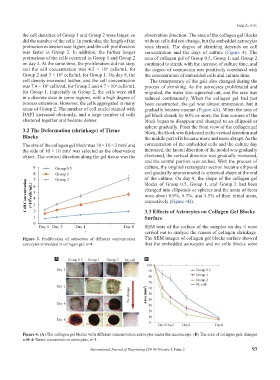
the cell densities of Group 1 and Group 2 were larger, so observation direction. The area of the collagen gel blocks
did the number of the cells. In particular, the length of the without cells did not change, but the embedded astrocytes
protrusion extension was higher, and the cell proliferation ones shrank. The degree of shrinking depends on cell
was faster in Group 2. In addition, the further longer concentration and the days of culture (Figure 4). The
protrusions of the cells occurred in Group 1 and Group 2 area of collagen gel of Group 0.5, Group 1, and Group 2
on day 4. At the same time, the proliferation did not stop, continued to shrink with the increase of culture time, and
and the cell concentration was 4.5 × 10 cells/mL for the degree of contraction was positively correlated with
6
Group 2 and 3 × 10 cells/mL for Group 1. On day 8, the the concentration of embedded cells and culture time.
6
cell density increased further, and the cell concentration The transparency of the gels also changed during the
was 7.4 × 10 cells/mL for Group 2 and 4.7 × 10 cells/mL process of shrinking. As the astrocytes proliferated and
6
6
for Group 1. Especially in Group 2, the cells were still migrated, the water was squeezed out, and the area was
in a discrete state in some regions, with a high degree of reduced continuously. When the collagen gel had just
process extension. However, the cells aggregated in many been constructed, the gel was almost transparent, but it
areas of Group 2. The number of cell nuclei stained with gradually became opaque (Figure 4A). When the area of
DAPI increased obviously, and a large number of cells gel block shrank by 60% or more, the four corners of the
clustered together and became denser. block began to disappear and changed to an ellipsoid or
sphere gradually. From the front view of the collagen gel
3.2 The Deformation (shrinkage) of Tissue block, the block was thickened in the vertical direction and
Blocks the middle part of it became more and more abrupt. As the
The size of the collagen gel block was 10 × 10 × 2 mm and concentration of the embedded cells and the culture day
3
the side of 10 × 10 mm was selected as the observation increased, the lateral direction of the model was gradually
2
object. The vertical direction along the gel tissue was the shortened, the vertical direction was gradually increased,
and the central portion was arched. With the process of
culture, the original rectangular section became ellipsoid
and gradually approximated to spherical shape at the end
of the culture. On day 8, the shape of the collagen gel
blocks of Group 0.5, Group 1, and Group 2 had been
changed into ellipsoids or spheres and the areas of them
were about 8.9%, 6.3%, and 4.5% of their initial areas,
respectively (Figure 4B).
3.3 Effects of Astrocytes on Collagen Gel Blocks
Surface
SEM tests of the surface of the samples on day 4 were
carried out to analyze the reason of collagen shrinkage.
Figure 3. Proliferation of astrocytes of different concentration The SEM images of collagen gel blocks surface showed
astrocytes embedded in collagen gel, n=4. that the embedded astrocytes and no cells blocks were
A B
Figure 4. (A) The collagen gel blocks with different concentration astrocytes under the microscope. (B) The area of collagen gels changes
with different concentration astrocytes, n=5.
International Journal of Bioprinting (2019)–Volume 5, Issue 2 93