
Page 100 - IJB-5-2
P. 100
In vitro model of the glial scar
and filling. Thus, the proposed 3D collagen gel tissue the increase of culture time. Tallinen et al. used a similar
[29]
constructed with astrocytes has scar formation properties, principle to construct a fold model of the human brain
and the glial scars are mainly produced by astrocytes. from a macro perspective. At this stage, the cells traction
It is quite coincidental that the similar phenomenon caused the deformation of the extracellular matrix [30-32] ,
of tissue shrinking also occurs in the brain of a Sorex resulting in a stronger collagen fiber aggregation.
araneus. Scientists have discovered that the skull of the With different initial cell concentrations, the degree of
Sorex araneus pup shrinks in the late summer, as well reduction is also different. The higher the concentration,
as the brain, whose weight is also reduced. During the the more cells proliferate and migrate, and the more
winter, the part of brain lost can grow back partly. This seriously the gel tissue shrinks. The collagen gel with
phenomenon is called Dehnel’s phenomenon [26-28] . It is astrocytes shrinks due to cell proliferation and migration.
similar to the results of this article, occurring in the brain. Consequently, the collagen fibers gradually extrude each
To a certain extent, it may provide some reference for other, the surface pores disappear, and the fiber pores
the study of brain plasticity and evolution. It may have a decrease. Meanwhile, the surface of the collagen gel is
certain value for reference to the study of brain plasticity deformed to form a relatively solid and wrinkled surface
and evolution. because of the movement of the cells. The process of gel
[33]
On the basis of this shrinkage, the folds can be found contraction is similar to the course of wound healing .
on the surface of the block, and the pores are reduced with The force produced by cell proliferation and migration
does not affect the collagen gel without cells. Hence, the
surface remains flat and porous. In addition, the shrinkage
rates of blocks with different concentration astrocytes
were relatively similar on day 8, but the moduli of them
were quite different. This indicates that the concentration
of cells also has a certain effect on the modulus when the
collagen gel shrinks. The higher the cell concentration,
the larger the collagen tissue modulus. From this, the
reason for collagen gel macroscopic and microscopic
size-changing can be inferred (Figure 10). The results
of the experiment can demonstrate that the model we
constructed can form glial scars.
5. Conclusion
In this study, a 3D astrocytes model with collagen
gel in vitro has been constructed. This model shows
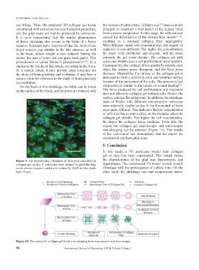
Figure 9. The hypertrophic cytoplasm of astrocytes embedded in the characteristics of the glial scar (hypertrophy and
collagen gel on day 8, astrocytes were stained by glial fibrillary hyperplasia). The constructed 3D model reveals overall
acidic protein in green, nuclei were stained by DAPI in blue, Scale shrinkage with the prolongation of culture time. On the
bars: 50 μm. other hand, the shrinkage rate and compression elastic
Figure 10. The reason for collagen gel block size changing from macroscopic and microscopic.
96 International Journal of Bioprinting (2019)–Volume 5, Issue 2