
Page 98 - IJB-5-2
P. 98
In vitro model of the glial scar
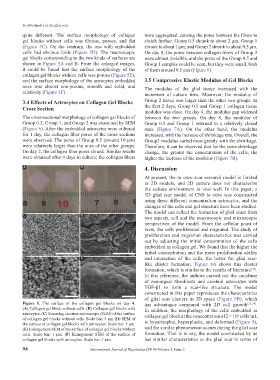
quite different. The surface morphology of collagen were aggregated, causing the pores between the fibers to
gel blocks without cells was fibrous, porous, and flat shrink further; Group 0.5 shrank to about 2 μm, Group 1
(Figure 5C). On the contrary, the one with embedded shrank to about 1 μm; and Group 2 shrank to about 0.5 μm.
cells had obvious folds (Figure 5D). The macroscopic On day 8, the pores between collagen fibers of Group 2
gel blocks corresponding to the two kinds of surfaces are were almost invisible, and the pores of the Group 0.5 and
shown in Figure 5A and B. From the enlarged images, Group 1 samples could be seen, but they were small, both
it could be found that the surface morphology of the of them around 0.5 μm (Figure 6).
collagen gel blocks without cells was porous (Figure 5E),
and the surface morphology of the astrocytes embedded 3.5 Compressive Elastic Modulus of Gel Blocks
ones was almost non-porous, smooth and solid, and The modulus of the glial tissue increased with the
relatively (Figure 5F). increment of culture time. Moerover, the modulus of
3.4 Effects of Astrocytes on Collagen Gel Blocks Group 2 tissue was larger than the other two groups. In
Cross Section the first 2 days, Group 0.5 and Group 1 collagen tissue
modulus was close. On day 4, the modulus gap widened
The cross-sectional morphology of collagen gel blocks of between the two groups. On day 8, the modulus of
Group 0.5, Group 1, and Group 2 was examined by SEM Group 0.5 and Group 1 returned to a relatively closed
(Figure 6). After the embedded astrocytes were cultured state (Figure 7A). On the other hand, the modulus
for 1 day, the collagen fiber pores of the cross sections increased, with the increase of shrinkage rate. Overall, the
were observed. The pores of Group 0.5 (around 10 μm) Group2 modulus varied most greatly with the shrinkage.
were relatively larger than the ones of the other groups. Therefore, it can be observed that for the same shrinkage
On day 2, the collagen fiber pores shrank. Similar results change, the greater the concentration of the cells, the
were obtained after 4 days in culture; the collagen fibers higher the increase of the modulus (Figure 7B).
A B 4. Discussion
At present, the in vitro scar research model is limited
to 2D models, and 2D culture does not characterize
the culture environment in vivo well. In this paper, a
3D glial scar model of CNS in vitro was constructed
using three different concentration astrocytes, and the
changes of the cells and gel structure have been studied.
The model can reflect the formation of glial scars from
C D
two aspects, cell and the macroscopic and microscopic
perspectives of the model. From the cellular point of
view, the cells proliferated and migrated. The study of
proliferation and migration characteristics was carried
out by adjusting the initial concentration of the cells
embedded in collagen gel. We found that the higher the
initial concentration and the more proliferation ability
E F and interaction of the cells, the better for glial scar-
like cluster formation. Figure 8A shows this cluster
formation, which is similar to the results of literature [20] .
In this reference, the authors carried out the coculture
of meningeal fibroblasts and cerebral astrocytes with
TGF-β1 to form a scar-like structure. The model
constructed in this paper reproduces the characteristics
of glial scar clusters in 3D space (Figure 8B), which
Figure 5. The surface of the collagen gel blocks on day 4. has advantages compared with 2D cell growth [23,24] .
(A) Collagen gel block without cells. (B) Collagen gel blocks with In addition, the morphology of the cells embedded in
astrocytes. (C) Scanning electron microscope (SEM) of the surface collagen gel block at the concentration of 2 × 10 cells/mL
6
of collagen gel blocks without cells. Scale bar: 5 μm (D) SEM of
the surface of collagen gel blocks with astrocytes. Scale bar: 5 μm. is hypertrophic, hyperplastic, and deformed (Figure 9),
(E) Enlargement SEM of the surface of collagen gel blocks without and the similar phenomenon occurs during the glial scar
cells. Scale bar: 1 μm. (F) Enlargement SEM of the surface of formation. That is to say, the model constructed by us
collagen gel blocks with astrocytes. Scale bar: 1 μm. has similar characteristics to the glial scar in terms of
94 International Journal of Bioprinting (2019)–Volume 5, Issue 2