
Page 132 - IJB-10-5
P. 132
International Journal of Bioprinting Liver printing: from structure to application
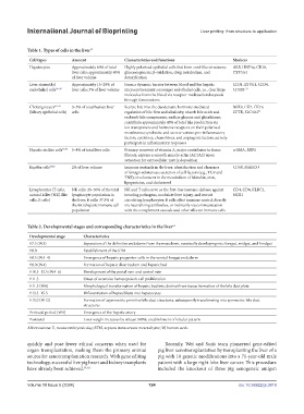
Table 1. Types of cells in the liver 35
Cell types Amount Characteristics and functions Markers
Hepatocytes Approximately 60% of total Highly polarized epithelial cells that form cord-like structures; ALB, HNF4α, CK18,
liver cells; approximately 80% gluconeogenesis, β-oxidation, drug metabolism, and CYP3A4
of liver volume detoxification
Liver sinusoidal Approximately 15–20% of Form a dynamic barrier between blood and the hepatic CD31, LYVE1, CD34,
endothelial cells 36–38 liver cells; 3% of liver volume microenvironment; scavenger endothelial cells, i.e., clear large CD105 39
molecules from the blood via receptor-mediated endocytosis
through fenestrations
Cholangiocytes 40–43 3–5% of total human liver Secrete bile into the duodenum; hormone-mediated SOX9, CK7, CK19,
(biliary epithelial cells) cells regulation of bile flow and alkalinity; absorb bile acids and CFTR, SLC4A2 44
reabsorb bile components, such as glucose and glutathione;
contribute approximately 40% of total bile production via
ion transporters and hormone receptors on their polarized
membranes; synthesize and secrete various pro-inflammatory
factors, cytokines, chemokines, and angiogenic factors; actively
participate in inflammatory responses
Hepatic stellate cells 45,46 5–8% of total liver cells Primary reservoir of vitamin A; major contributor to tissue α-SMA, RBP1
fibrosis; express α-smooth muscle actin (ACTA2) upon
activation for extracellular matrix deposition
Kupffer cells 47,48 2% of liver volume Immune sentinels in the liver; identification and clearance CD68, MARCO
of foreign substances; secretion of cell factors (e.g., TGF and
TNF); involvement in the metabolism of bilirubin, iron,
lipoproteins, and cholesterol
Lymphocytes (T cells, NK cells: 20–30% of the total NK and T cells serve as the first-line immune defense against CD4, CD8, KLRC1,
natural killer [NK]-like lymphocyte population in invading pathogens, modulate liver injury, and recruit NCR1
cells, B cells) the liver; B cells: 37.5% of circulating lymphocytes; B cells affect immune control directly
the intrahepatic immune cell via neutralizing antibodies, or indirectly via communication
population with the complement cascade and other effector immune cells
Table 2. Developmental stages and corresponding characteristics in the liver 52
Developmental stage Characteristics
E7.5 (W3) Separation of the definitive endoderm from the mesoderm, eventually developing into foregut, midgut, and hindgut
E8.0 Establishment of the STM
E8.5 (W3–4) Emergence of hepatic progenitor cells in the ventral foregut endoderm
E9.0 (W4) Formation of hepatic diverticulum and hepatic bud
E10.5–12.5 (W4–6) Development of the portal vein and central vein
E11.5 Onset of extensive hematopoietic cell proliferation
E11.5 (W6) Morphological transformation of hepatic bud into distinct liver tissue; formation of the bile duct plate
E13.5–15.5 Differentiation of hepatoblasts into hepatocytes
E15.0 (W12) Formation of asymmetric primitive bile duct structures, subsequently transforming into symmetric bile duct
structures
Perinatal period (W8) Emergence of the hepatic artery
Postnatal Liver weight increases by at least 500%; establishment of lobular pattern
Abbreviations: E, mouse embryonic day; STM, septum transversum mesenchyme; W, human week.
quickly and pose fewer ethical concerns when used for Recently, Wei and Sun’s team pioneered gene-edited
organ transplantation, making them the primary animal pig liver xenotransplantation by transplanting the liver of a
source for xenotransplantation research. With gene editing pig with 10 genetic modifications into a 71-year-old male
technology, successful live pig heart and kidney transplants patient with a large right lobe liver cancer. This procedure
have already been achieved. 61,62 included the knockout of three pig xenogeneic antigen
Volume 10 Issue 5 (2024) 124 doi: 10.36922/ijb.3819