
Page 195 - IJB-10-5
P. 195
International Journal of Bioprinting Structural design of D-surface scaffolds
scaffold with surrounding tissues was examined by micro- to edge. In contrast, the wall thickness of the cube sample
CT and histological analysis. The implanted scaffold with became thicker closer to the bottom. As the thickness
tissues was fixed overnight with 4% paraformaldehyde increased, the porous size became smaller. The successful
and scanned using micro-CT (Xradia 610, 70 kV, 8.5 W; FFF-printed D-surfaces suggested the printability of PBAT/
Zeiss, Germany). The fixed scaffolds were then embedded PLA into fine lattice structures. Good ductility of PBAT/PLA
in paraffin, sliced in 4-µm thickness, and stained by has been demonstrated with an ultimate fracture strength
hematoxylin-eosin (H&E). All protocols were approved by and elongation at break of 19 MPa and 350%, respectively. 19
the Institutional Animal Ethical Committee (IAEC) of The Since FFF is a layer-by-layer deposition technique, the
Second Affiliated Hospital of Dalian Medical University printing quality is important for the structures’ mechanical
(approval no: AEE22048). All methods were performed in performance. The internal 3D structure of D-surfaces was
accordance with relevant guidelines and regulations. observed by CT. Figure 3 features the reconstruction and
2.8. Statistical analysis slicing images of uniform and graded D-surfaces (0.8–1.6
Quantitative data are presented as the mean ± standard mm) in the x–z plane. In Figure 3a, the D-surface sample
deviation (SD). One-way Analysis of Variance (ANOVA) has a thickness of 1.2 mm. The homogeneous D-surface
and subsequent comparisons between each group of displayed uniform thickness, and the cellular topology was
scaffolds were analyzed using SPSS 25.0 statistics software. confirmed via sliced images. From the sliced images, the
D-surface exhibited a continuous 3D lattice structure with
3. Results and discussion no obvious manufacturing defects, making it beneficial for
load-carrying and stress transfer. Besides, the interconnected
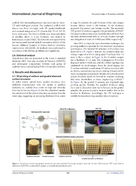
3.1. 3D printing of uniform and graded diamond porous structure would be favored for nutrient exchange
surface structures and waste metabolism as tissue engineering scaffolds.
30
To better mimic natural bone, graded structures were In Figure 3b, the graded D-surfaces also displayed high
additively manufactured with two modes of gradient printing quality. Notably, the gradient thickness along
thickness, i.e., radially from center to edge and vertically the z-axis is distinctive from top to bottom. In the graded
from top to bottom (Figure 2). For the cylindrical sample, samples, the porous structure became smaller due to the
the central part of the porous structure was sparser than the increase in thickness. Accordingly, the FFF technique is
outer edge, suggesting an increasing thickness from center suitable for manufacturing complex lattice structures.
Figure 2. 3D-printed uniform and graded cylindrical and cubic diamond (D)-surface samples. Scale bar: 10 mm. Abbreviations: FFF: fused filament
fabrication; PBAT/PLA: poly(butylene adipate-co-terephthalate)/poly(lactic acid).
Volume 10 Issue 5 (2024) 187 doi: 10.36922/ijb.3416