
Page 200 - IJB-10-5
P. 200
International Journal of Bioprinting Structural design of D-surface scaffolds
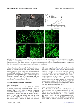
Figure 9. The in vitro biological performance of D-surface scaffolds. (a) Microstructure of the samples obtained with scanning electron microscopy (SEM).
(b) Cell Counting Kit-8 (CCK-8) assay results of the L929 culture. (c and d) Fluorescence imaging of (c) the poly(butylene adipate-co-terephthalate)/
poly(lactic acid) (PBAT/PLA) scaffolds and (d) PBAT/PLA with platelet-rich plasma (PBAT/PLA+PRP) scaffolds proliferated for 24, 48, and 96 h. Scale
bars: (a) 500 µm; (c and d) 100 µm. Magnification: 200×. Abbreviation: OD: optical density.
different from the uniform sample. The large deformation OD value, suggesting enhanced cell proliferation with
tended to concentrate on the region with thinner prolonged culture time (Figure 9b). Cell proliferation on
thickness, and the thicker bottom part well supported the the PBAT/PLA and PBAT/PLA+PRP scaffolds was then
external load, correlating to the enhanced compression observed through fluorescence staining. After 24 h of
performance of the graded samples. Therefore, the graded cell culture, the live cells were attached to the scaffolds,
D-surface structures have a good load-carrying and exhibiting a clear porous structure. As the cell culture
energy absorption capacity, making them suitable as bone continued, the amount of live cells sharply increased, and
implants and tissue engineering scaffolds. no PI-stained dead cells were detected. The cell viability
assay demonstrated that both the PBAT/PLA and PBAT/
3.3. In vitro biocompatibility of scaffolds PLA+PRP scaffolds possess good biocompatibility, and the
PRP layer promoted cell proliferation. 32
3.3.1. Cell viability
The surface of the bare and the PRP-loaded (PBAT/ 3.3.2. Mineralization activity
PLA+PRP) scaffolds was observed by SEM. It could be The ALP activity of the 3D-printed scaffold groups was
observed that the PBAT/PLA+PRP scaffold was coated further evaluated, and the results are displayed in Figure 10.
with a thin PRP layer (Figure 9a). The viability of L929 ALP is an early-stage indicator to assess osteoblast
fibroblasts was assayed by comparing between the PRP- differentiation. As displayed, ALP activity increased
loaded scaffold, the bare scaffold, and the cell plate with longer culture time. The stimulation of osteoblast
(control). The CCK-8 assay results demonstrated that differentiation was evident in the PRP-loaded scaffold
the PBAT/PLA+PRP scaffold exhibited an enhanced group (PBAT/PLA+PRP), indicating that PRP loading
Volume 10 Issue 5 (2024) 192 doi: 10.36922/ijb.3416