Page 201 - IJB-10-5
P. 201
International Journal of Bioprinting                                 Structural design of D-surface scaffolds


                                                               could accelerate mineralization via released cytokines in
                                                               the osteogenic maturation process. 33

                                                               3.4. In vivo biocompatibility of scaffolds
                                                               Based on the in vitro biocompatibility, it is critical to
                                                               investigate the bone repair effect of PRP-loaded D-surface
                                                               scaffolds in a pre-clinical setting. A rabbit femur defect
                                                               model was created using New Zealand white rabbits.
                                                               The PRP-loaded scaffolds and the operation process are
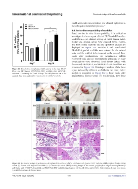
                                                               displayed in  Figure 11a. PBAT/PLA and PRP-loaded
                                                               PBAT/PLA graded scaffolds were selected for the animal
                                                               tests, and the unfilled defect was set as the control. Four
                                                               weeks after implantation, the experimental rabbits
                                                               recovered well, and no postoperative infection or other
                                                               complications were observed. Local femur defects with
                                                               the control, PBAT/PLA, and PBAT/PLA+PRP scaffolds are
                                                               presented in Figure 11b. Histological analysis of the bone
            Figure 10. The alkaline phosphatase (ALP) activity in the bare (PBAT/  repair induced by different scaffolds for the rabbit defect
            PLA) and PRP-loaded (PBAT/PLA+PRP) scaffolds after MC3T3-E1
            subclone 14 culturing for 7 and 14 days. The cell plate was set as the   models is presented in  Figure 11c–e. Four weeks after
            control. Data were analyzed by t-test (n = 3). *p < 0.05; **p < 0.01.   implantation, fibrous tissue (F) proliferation, new bone


            Figure 11. The in vivo biological performance of implanted D-surface scaffolds. (a) Platelet-rich plasma (PRP)-loaded scaffolds implanted in the rabbit
            defect. (b) Femurs with implanted scaffolds. (c–e) Hematoxylin-eosin (H&E) staining images of the control, poly(butylene adipate-co-terephthalate)/
            poly(lactic acid) (PBAT/PLA) scaffold, and PBAT/PLA+PRP scaffold. Magnification: (c) 40×, (d) 100×, and (e) 400×. Abbreviations: B: new bone tissue;
            S: scaffolds; E: edema; F: fibrous tissue.

            Volume 10 Issue 5 (2024)                       193                                doi: 10.36922/ijb.3416

