
Page 202 - IJB-10-5
P. 202
International Journal of Bioprinting Structural design of D-surface scaffolds
(B) formation, and mild edema (E) were observed among bioactivity factors to foster an early-stage conducive 3D
the three groups of bone cortex. It was noticeable that microenvironment for osteoblasts.
34
the PBAT/PLA+PRP scaffold has a much higher degree of
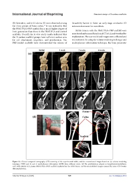
bone generation than those in the PBAT/PLA and control Rabbit femurs with the PBAT/PLA+PRP scaffold were
scaffolds. Overall, the in vitro study results indicated that monitored and scanned by a clinical CT at 1, 2, and 4 weeks after
the D-surface scaffold groups have sufficient surface area implantation. The scanned sliced images were collected and
for cell attachment, migration, and proliferation. The reconstituted. By using the volume rendering technique and
PRP-loaded scaffolds have demonstrated the release of multi-planner reformation technique, the bone parameter
Figure 12. Clinical computed tomography (CT) scanning of the experimental rabbit, and the reconstituted images based on (a) volume rendering
technique (VRT) and (b and c) multi-planner reformation (MPR) from different views. (d) The poly(butylene adipate-co-terephthalate)/poly(lactic
acid) with platelet-rich plasma (PBAT/PLA+PRP) scaffold implanted in the rabbit femur, and the reconstituted images scanned by micro-CT 4 weeks
after implantation.
Volume 10 Issue 5 (2024) 194 doi: 10.36922/ijb.3416