
Page 203 - IJB-10-5
P. 203
International Journal of Bioprinting Structural design of D-surface scaffolds
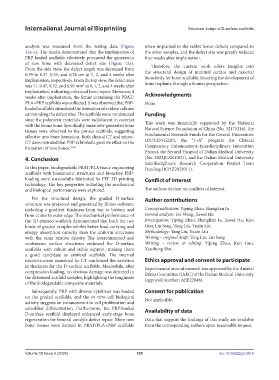
analysis was measured from the testing data (Figure when implanted in the rabbit femur defects compared to
12a–c). The results demonstrated that the implantation of the other samples, and the defect size was greatly reduced
PRP-loaded scaffolds effectively promoted the generation four weeks after implantation.
of new bone with decreased defect size (Figure 12a). Therefore, the current work offers insights into
From the side view, the defect length was decreased from the structural design of minimal surface and material
0.39 to 0.37, 0.35, and 0.28 cm at 1, 2, and 4 weeks after bioactivity for bone scaffolds, fostering the development of
implantation, respectively. From the top view, the defect area
was 11, 9.47, 8.32, and 6.99 mm at 0, 1, 2, and 4 weeks after bone implants through a bionics perspective.
2
implantation, indicating enhanced bone repair. Moreover, 4 Acknowledgments
weeks after implantation, the femur containing the PBAT/
PLA+PRP scaffolds was collected. It was observed that PRP- None.
loaded scaffolds stimulated the formation of evident calluses
surrounding the defect sites. The scaffolds were not detected Funding
since the polymeric materials were radiolucent in contrast This work was financially supported by the National
with the femur bone. Specifically, some new-generated bone
tissues were observed in the porous scaffolds, suggesting Natural Science Foundation of China (No. 52175216), the
effective new bone formation. Both clinical CT and micro- Fundamental Research Funds for the Central Universities
CT demonstrated that PRP exhibited a positive effect on the (DUT23YG220), the “1+X” program for Clinical
formation of new bones. 35,36 Competency Enhancement–Interdisciplinary Innovation
Project, the Second Hospital of Dalian Medical University
4. Conclusion (No. 2022JCXKYB21), and the Dalian Medical University
Interdisciplinary Research Cooperation Project Team
In this paper, biodegradable PBAT/PLA tissue engineering Funding (JCHZ2023011).
scaffolds with biomimetic structures and bioactive PRP-
loading were successfully fabricated by FFF 3D printing Conflict of interest
technology. The key properties including the mechanical
and biological performance were explored. The authors declare no conflicts of interest.
For the structural design, the graded D-surface Author contributions
structure was proposed and generated by Rhino software,
including a gradient thickness from top to bottom and Conceptualization: Yiping Zhao, Shanglian Ju
from center to outer edge. The mechanical performance of Formal analysis: Fei Wang, Jiawei Hu
the 3D-printed scaffolds demonstrated that both the two Investigation: Yiping Zhao, Shanglian Ju, Jiawei Hu, Kun
kinds of graded samples exhibit better load-carrying and Guo, Lin Sang, Tang Liu, Yuxin Lin
energy absorption capacity than the uniform structures Methodology: Tang Liu, Yuxin Liu
with the same relative density. The interconnected and Writing – original draft: Tang Liu, Lin Sang
continuous surface structures endowed the D-surface Writing – review & editing: Yiping Zhao, Kun Guo,
scaffolds with robust and stable support, making them Xiaohong Shu
a good candidate as artificial scaffolds. The internal
microstructure examined by CT confirmed the variation Ethics approval and consent to participate
in thickness for the D-surface scaffolds. Meanwhile, after Experimental animal research was approved by the Animal
compression loading, no obvious damage was detected in Ethics Committee (IAEC) of the Dalian Medical University
the deformed scaffold samples, highlighting the toughness (approval number: AEE22048).
of the biodegradable composite materials.
Subsequently, PRP with diverse cytokines was loaded Consent for publication
on the graded scaffolds, and the in vitro cell biological Not applicable.
activity suggests an enhancement in cell proliferation and
osteoblast differentiation. Furthermore, the PRP-loaded Availability of data
D-surface scaffold displayed enhanced early-stage bone
regeneration for femoral condyle defect repair. More new Data that support the findings of this study are available
bone tissues were formed in PBAT/PLA+PRP scaffolds from the corresponding authors upon reasonable request.
Volume 10 Issue 5 (2024) 195 doi: 10.36922/ijb.3416