
Page 212 - IJB-10-5
P. 212
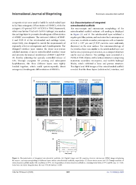
International Journal of Bioprinting                                     Biomimetic osteochondral scaffold


            composite struts were used to build the subchondral layer   3.2. Characterization of integrated
            to facilitate osteogenic differentiation of rBMSCs, while the   osteochondral scaffolds
            cryogenic 3D-printed FGF-18/P(DLLA-TMC) framework,   The macroscopic and microscopic morphology of the
            which was further filled with GelMA hydrogel, was used as   osteochondral scaffold without cell seeding is displayed
            the cartilage layer to promote chondrogenic differentiation   in  Figure 2A and  B. The subchondral layer exhibited a
            of rBMSC microspheres. The sustained delivery of BMP-  regular grid-like pattern, and each strut had a microporous
            2  and FGF-18  at  the  subchondral and  cartilage  layers,   structure, in which secondary micropores with a diameter
            respectively, was designed to match the requirements of   of 8.10 ± 1.87 μm and  β-TCP particles were uniformly
            regionally effective osteogenesis and chondrogenesis. The   dispersed on the strut surface. The micromorphology of
            designed interface layer mimics the dense non-porous   the interface layer was similar to the subchondral layer and
            calcified anatomy of native osteochondral interface tissue   had no macroporous grid structure, as a compact structure
            and prevents the mutual interference of BMP-2 and FGF-  can be used as a barrier. The cartilage layer consisted of a
            18, thereby achieving the spatially controlled release of   P(DLLA-TMC) frame, with a latticed structure comprising
            GFs. Through cryogenic 3D printing and subsequent   numerous secondary micropores, and GelMA hydrogel
            lyophilization, the  three different layers  were tightly   blocks, which exhibited a loose and porous structure.
            bonded together, which could spatiotemporally direct   The digital and SEM images of the osteochondral scaffold
            osteogenic/chondrogenic differentiation of rBMSCs.  revealed that the three layers (subchondral, interface, and


            Figure 2. Characterization of integrated osteochondral scaffolds. (A) Digital images and (B) scanning electron microscope (SEM) images displaying
            the macro- and micromorphology of different layers within osteochondral scaffolds, respectively. (C) Stress–strain curves and (D) Young’s modulus of
            osteochondral scaffolds under wet conditions at 37°C. (E) Degradation of osteochondral scaffolds in a shaking water bath at 37°C and 80 rpm. *p < 0.05.
            Scale bars: (A) XXX. Magnification: (B, top) ×50; (B, bottom) ×1000.


            Volume 10 Issue 5 (2024)                       204                                doi: 10.36922/ijb.3229