
Page 216 - IJB-10-5
P. 216
International Journal of Bioprinting Biomimetic osteochondral scaffold
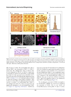
Figure 5. Fabrication of rat bone marrow mesenchymal stem cell (rBMSC) microspheres. (A) rBMSCs and rBMSC microspheres that are (i) seeded, (ii)
cultivated for 24 h, and (iii) removed in/from the microwell culture plate. (B) Histograms of diameter distribution of rBMSC microspheres. (C) Scanning
electron microscope (SEM) images of rBMSC microspheres after lyophilization. (D) Immunofluorescence staining of F-actin (red) and DAPI (blue) in
rBMSC microspheres. (E) The inspiration and structural similarity between cartilage lacunae and rBMSC microspheres. Magnification: ×100 (left) and
×400 (right) . Abbreviation: GelMA, gelatin methacrylate.
after 14 days of culture. The subchondral layer with or there was scarcely any ALP expression in the cartilage layer
without BMP-2 was designated as BMP-2 or BMP-2 , (FGF-18 ). Furthermore, gene expression analysis revealed
−
+
+
respectively. The cartilage layer with or without FGF- that the expression levels of RUNX2, COL I, and OCN in
−
+
18 was designated as FGF-18 or FGF-18 , respectively. the subchondral layer (BMP-2 ) were significantly higher
−
−
Compared to the subchondral layer (BMP-2 ) and cartilage compared to the tissue culture plate group but significantly
layer (FGF-18 ), the expression levels of RUNX2, COL I, lower compared to the subchondral layer (BMP-2 ). In
+
+
and OCN were more pronounced in the subchondral layer addition, there was no significant difference in RUNX2,
(BMP-2 ) (Figure 7A–C). ALP staining was also conducted COL I, and OCN expression levels in the subchondral
+
to reflect the osteogenic differentiation of rBMSCs. After layer (BMP-2 ) and OM group (Figure 7D).
+
14 days of culture, there was minimal expression of ALP
(stained in purple) in the subchondral layer (BMP-2 ) The chondrogenic potential of rBMSC microspheres
−
and cartilage layer (FGF-18 ), while ALP expression was in osteochondral scaffolds was assessed by visualizing
+
significantly upregulated in the subchondral layer (BMP- the expression of SOX9, COL II, and ACAN via
2 ) (Figure S1, Supporting Information). Additionally, immunofluorescence staining after 21 days of culture.
+
Volume 10 Issue 5 (2024) 208 doi: 10.36922/ijb.3229