
Page 214 - IJB-10-5
P. 214
International Journal of Bioprinting Biomimetic osteochondral scaffold
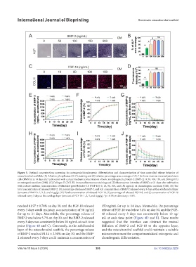
Figure 3. Optimal concentrations screening for osteogenic/chondrogenic differentiation and characterization of time-controlled release behavior of
osteochondral scaffolds. (A) Alkaline phosphatase (ALP) staining and (B) relative percentage area coverage of ALP for bone marrow mesenchymal stem
cells (BMSCs) at 14 days after cultivation with culture medium (concentration of bone morphogenetic protein-2 [BMP-2]: 0, 50, 100, 150, and 200 ng/mL)
or osteogenic medium (OM). (C) Collagen II (COL II) immunofluorescence staining and (D) fluorescence intensity of BMSCs at 21 days after cultivation
with culture medium (concentration of fibroblast growth factor-18 [FGF-18]: 0, 10, 50, 100, and 150 ng/mL) or chondrogenic medium (CM). (E) The
total concentration of released BMP-2, (F) percentage of released BMP-2, and (G) concentration of BMP-2 released every 3 days at the subchondral layer
(amount of BMP-2: 1, 2, 3, and 4 μg/g). (H) Total concentration of released FGF-18, (I) percentage of released FGF-18, and (J) concentration of FGF-18
released every 3 days at the cartilage layer (amount of FGF-18: 1, 2, 3, and 4 μg/g). *p < 0.05; ns denotes p > 0.05.
reached 61.97 ± 0.76% on day 30, and the FGF-18 released 150 ng/mL for up to 14 days. Meanwhile, the percentage
every 3 days could maintain a concentration of 50 ng/mL release of FGF-18 was below 5.4% on day 30, and the FGF-
for up to 21 days. Meanwhile, the percentage release of 18 released every 3 days was consistently below 10 ng/
BMP-2 was below 5.7% on day 30, and the BMP-2 released mL at each time point (Figure 4D and E). These results
every 3 days was consistently below 20 ng/mL at each time suggested that the interface can obstruct the mutual
point (Figure 4B and C). Conversely, in the subchondral diffusion of BMP-2 and FGF-18 to the opposite layer,
layer of the osteochondral scaffold, the percentage release and the osteochondral scaffold could maintain a suitable
of BMP-2 reached 81.14 ± 2.30% on day 30, and the BMP- microenvironment for compartmentalized osteogenic and
2 released every 3 days could maintain a concentration of chondrogenic differentiation.
Volume 10 Issue 5 (2024) 206 doi: 10.36922/ijb.3229