
Page 446 - IJB-10-5
P. 446
International Journal of Bioprinting Bioprinting for large-sized tissue delivery
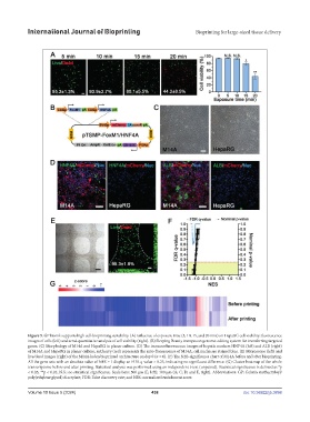
Figure 5. GP bioink supports high cell-bioprinting suitability. (A) Influence of exposure time (5, 10, 15, and 20 min) on HepaRG cell viability: fluorescence
images of cells (left) and semi-quantitative analysis of cell viability (right). (B) Sleeping Beauty transposon genome-editing system for transferring targeted
genes. (C) Morphology of M14A and HepaRG in planar culture. (D) The immunofluorescence images of hepatic markers HNF4A (left) and ALB (right)
of M14A and HepaRG in planar culture; mCherry (red) represents the auto-fluorescence of M14A; cell nuclei are stained blue. (E) Microscope (left) and
live/dead images (right) of the M14A-laden bioprinted architecture on day 0 (n = 6). (F) The NES-significance chart of M14A before and after bioprinting.
All the gene sets with an absolute value of NES ≥ 1 display an FDR q-value > 0.25, indicating no significant difference. (G) Cluster heatmap of the whole
transcriptome before and after printing. Statistical analysis was performed using an independent t-test (unpaired). Statistical significance is defined as *p
< 0.05; **p < 0.01; N.S.: no statistical significance. Scale bars: 500 μm (E, left); 100 μm (A; C; D; and E, right). Abbreviations: GP: Gelatin methacryloyl/
poly(ethylene glycol) diacrylate; FDR: False discovery rate; and NES: normalized enrichment score.
Volume 10 Issue 5 (2024) 438 doi: 10.36922/ijb.3898