
Page 490 - IJB-10-5
P. 490
International Journal of Bioprinting Biocompatible 3D-printed radiotherapy spacer
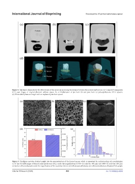
Figure 4. This figure demonstrates the effectiveness of the spacer in increasing the distance between the prostate and rectum. (a) Computed tomography
(CT) scan images of original phantom without spacer. (b, c) Implantation of pre-foam (b) and post-foam (c) polycaprolactone (PCL) spacers.
(d) 3D modeled phantom images with an implanted post-foam spacer.
Figure 5. This figure provides detailed insight into the microstructure of the foamed spacer, which is important for understanding cell morphologies.
(a, b) The FE-SEM images of foamed polycaprolactone (PCL) under the magnifications of 500× (a; scale bar: 300 µm) and 1000× (b; scale bar: 100 µm).
(c) Section of foamed sample under the magnification of 70× (scale bar: 2mm). (d) Cell size and cell density. (e) Cell size distribution of foamed PCL sample.
Volume 10 Issue 5 (2024) 482 doi: 10.36922/ijb.4252