
Page 487 - IJB-10-5
P. 487
International Journal of Bioprinting Biocompatible 3D-printed radiotherapy spacer
based 3D-printed, microcellular foamed spacer for prostate Table 1. Experimental parameters of microcellular foaming
cancer RT. process
2. Materials and methods Property Value
Saturation pressure (MPa) 8 ± 0.1
2.1. Materials Saturation temperature (°C) 40 ± 1
A PCL filament (eMate, eSUN, China) was prepared as Saturation time (min) 15
the polymer matrix for the fabrication of spacers using 3D
printing and MCP. PCL is a semi-crystalline polymer with Depressurization ratio (MPa/s) 0.3
a density of 1.16 g/cm , a glass transition temperature (T )
3
g
of −60°C, and a melting temperature (T ) of 60°C. PCL is detailed 3D printing parameters for PCL included a nozzle
m
readily available as a commercial or medical filament and temperature of 170°C and a bed temperature of 40°C.
has the advantage of being compatible with standard 3D The nozzle diameter was set to 0.4 mm, with a printing
printers. CO (purity: 99.9%; 40 L; Samheung GasTech, speed of 15 mm/s. The layer thickness was 0.25 mm. The
2
Seoul, Republic of Korea) was used as a blowing agent in G-code was generated using a 3D printing open-source
the MCP to achieve stable saturation and high solubility. slicing program (Cura slicer program, Geldermalsen,
Additionally, scCO was selected because of its sterilization the Netherlands). The infill pattern was set to a 50% grid
2
capability, making CO a suitable blowing agent. pattern. 3D modeling was performed using a computer-
2
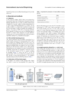
2.2. Microcellular foaming process aided design (CAD) software (Creo 4.0, PTC Inc., USA).
The spacer printed by the 3D printer was placed in a Before designing the spacer, preliminary experiments were
batch chamber, where scCO2 was generated at 40°C and 8 conducted to assess the volume expansion rate and cell
MPa, to allow CO diffusion into the PCL for 15 min. For morphology induced by the MCP. For these experiments,
2
3D-printed samples with 50% infill, we set the thickness of samples with dimensions of 20 mm × 20 mm × 5 mm
filaments extruded from the nozzle to less than 0.4 mm, were printed.
allowing for full saturation within 15 min. Subsequently, 2.4. Length expansion along the x-, y-, and z-axes
depressurization was employed to induce thermodynamic After placing the 3D-printed spacer in a batch chamber,
instability, causing the CO dissolved in the free volume scCO was diffused into the spacer for 15 min. Once the
2
of the amorphous regions of PCL to expand, resulting foaming process was completed through depressurization,
2
in foaming (Figure 1). Given that the T of PCL is the spacer achieved its final planned size and shape. Upon
g
approximately −60°C, microcellular foam is formed inside
the chamber during depressurization. 24–26 The detailed the completion of depressurization, the volume of the
experimental parameters for MCP are listed in Table 1. spacer expanded. The expanded spacer was then accurately
positioned between the rectum and the prostate during the
2.3. Fabrication of 3D-printed sample procedure, ensuring a separation of >10 mm between the
A 3D printer (X1-Carbon Combo; Bambu Lab, China) two organs. After completion of the volume expansion using
was used to create the biocompatible PCL spacer. The the MCP, the length expansion ratio (%) was measured to
Figure 1. Schematic of the overall microcellular foaming process.
Volume 10 Issue 5 (2024) 479 doi: 10.36922/ijb.4252