
Page 85 - IJB-10-5
P. 85
International Journal of Bioprinting dECM bioink for 3D musculoskeletal tissue reg.
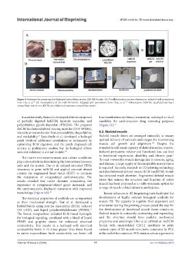
Figure 4. Strategies for preparing decellularized extracellular matrix (dECM) bioinks. (A) Decellularization process of meniscus. Adapted with permission
134
38
from Das et al. (B) Development of the hdECM bioink. Adapted with permission from Chae et al. Abbreviation: hdECM, decellularized heart
extracellular matrix; me-dECM, decellularized meniscus extracellular matrix.
In another study, Shin et al. developed a bioink composed functionalization and tissue maturation, making it an ideal
of partially digested hdECM, laponite nanoclay, and candidate for cardiovascular drug screening purposes
poly(ethylene glycol)-diacrylate (PEGDA). The prepared (Figure 5E). 41
dECM biochain exhibited varying modulus (13.4–89 kPa),
viscosity at rest and under flow, extrudability, shape fidelity, 6.2. Skeletal muscle
and stackability. Sanz-Fraile et al. developed a hydrogel Skeletal muscle fibers are arranged uniaxially to ensure
27
patch (without additional crosslinkers or enhancers) by optimal delivery of nutrients and oxygen for maintaining
150
optimizing ECM digestion, and the patch displayed cell muscle cell growth and alignment. Despite the
activity in preliminary studies, but its biological effects remarkable self-repair capacity of skeletal muscles, trauma-
were not validated in animal models. 149 induced permanent volume and functional loss can lead
to functional impairment, disability, and chronic pain.
8
The matrix microenvironment and culture conditions
play a pivotal role in determining the interactions between To treat irreversible muscle damage due to exercise, aging,
and disease, a large supply of biocompatible muscle tissue
cells and the matrix. Das et al. utilized extruded PEVA
structures to print hdECM and applied external stimuli is required. Recently, research on 3D printing technology
outside the engineered heart tissue (EHT) to promote and decellularized skeletal muscle ECM (mdECM) bioink
the maturation of encapsulated cardiomyocytes. The has attracted much attention. Engineered skeletal muscle
results revealed that under dynamic stimulation, the tissue that mimics the structure and function of native
expression of myogenesis-related genes increased, and muscle has been proposed as a viable treatment option for
the cardiomyocytes displayed maturation with improved a range of muscle-related ailments and injuries.
morphology (Figure 5D). 38 Recent advances in 3D bioprinting have facilitated the
The electrical properties of scaffolds are as important development of highly ordered biological scaffolds for
as their mechanical strength. Tsui et al. developed a muscle TE. The capacity to regulate fiber alignment and
hybrid bioink using porcine myocardial dECM, reduced orientation during the printing process paved the way for
GO (rGO), and liquid polydimethylsiloxane (PDMS). the development of functional muscle tissue constructs.
41
The bioink composition included ECM-based hydrogels Skeletal muscle is constantly contracting and expanding,
for biological signaling, combined with a blend of liquid and the structure should have realistic mechanical
PDMS and graphite sheets for enhanced electrical properties and anisotropy. Choi et al. utilized an mdECM
conductivity. This results in a hydrogel with electrical bioink with encapsulated C2C12 myoblasts to print
conductivity levels 3–10 times greater than those found various types of 3D muscle structures, constraint by PCL
in native myocardium. Such conductivity can foster cell at the end of the construct. PCL tension acts as a geometric
Volume 10 Issue 5 (2024) 77 doi: 10.36922/ijb.3418