
Page 46 - IJB-6-2
P. 46
3D freeform printing of nanocomposite hydrogels
A B
C D
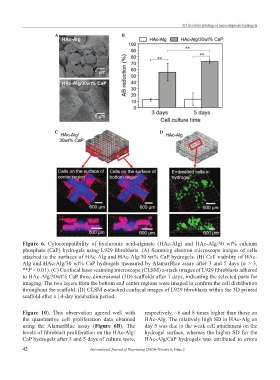
Figure 6. Cytocompatibility of hyaluronic acid-alginate (HAc-Alg) and HAc-Alg/30 wt% calcium
phosphate (CaP) hydrogels using L929 fibroblasts. (A) Scanning electron microscope images of cells
attached to the surfaces of HAc-Alg and HAc-Alg/30 wt% CaP hydrogels. (B) Cell viability of HAc-
Alg and HAc-Alg/30 wt% CaP hydrogels measured by AlamarBlue assay after 3 and 5 days (n > 3,
**P < 0.01). (C) Confocal laser scanning microscope (CLSM) z-stack images of L929 fibroblasts adhered
to HAc-Alg/30wt% CaP three-dimensional (3D) scaffolds after 7 days, indicating the selected parts for
imaging. The two layers from the bottom and center regions were imaged to confirm the cell distribution
throughout the scaffold. (D) CLSM z-stacked confocal images of L929 fibroblasts within the 3D printed
scaffold after a 14-day incubation period.
Figure 10). This observation agreed well with respectively, ~6 and 8 times higher than those on
the quantitative cell proliferation data obtained HAc-Alg. The relatively high SD in HAc-Alg on
using the AlamarBlue assay (Figure 6B). The day 5 was due to the weak cell attachment on the
levels of fibroblast proliferation on the HAc-Alg/ hydrogel surface, whereas the higher SD for the
CaP hydrogels after 3 and 5 days of culture were, HAc-Alg/CaP hydrogels was attributed to errors
42 International Journal of Bioprinting (2020)–Volume 6, Issue 2