
Page 48 - IJB-6-2
P. 48
3D freeform printing of nanocomposite hydrogels
A B
C D
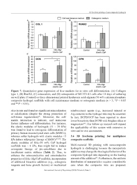
Figure 7. Quantitative gene expression of four markers for in vitro cell differentiation. (A) Collagen
type 1, (B) RunX2, (C) osteocalcin, and (D) osteopontin of MC3T3-E1 cells after 14 days of culturing
on well plate (Control) or three-dimensional printed hyaluronic acid-alginate/30 wt% calcium phosphate
composite hydrogel scaffolds with cell maintenance medium or osteogenic medium (n > 3, *P < 0.05
and **P < 0.01).
sites in rats and found no significant mineralization reinforcement agents (e.g., increased mineral or
or calcification (despite the strong promotion of Alg contents) to the hydrogel inks may be essential.
soft-tissue regeneration) . Moreover, the cell/ In fact, DCPD/OCP has been reported to show
[5]
matrix interaction is intricate, and numerous lower bioactivity than DCPD with bioglass silica or
factors influence cell differentiation. For instance, magnesium . Our follow-up research will expand
[45]
the elastic modulus of hydrogels (11 – 30 kPa) the applicability of this system with extensive in
was found to lead to osteogenic differentiation of vitro and in vivo assessments.
primary human mesenchymal stem cells (hMSCs),
whereas softer hydrogels with elastic modulus <5 3.6 3D freeform printing for multiphase
kPa induce adipogenic lineage of hMSCs [43,44] . The composite scaffolds
elastic modulus of HAc-30 wt% CaP hydrogel
scaffolds was ~ 6 kPa, thus might fail to induce Multi-material 3D printing with nanocomposite
osteogenic lineage of pre-osteoblasts due to hydrogels is challenging because the nanoparticle
insufficient matrix stiffness (Table 2). Thus, to additives may change the rheological behavior of the
enhance the osteoconductive and osteoinductive composite hydrogel inks depending on the loading
properties of HAc-Alg/CaP scaffolds, incorporation amount of the additives . Furthermore, the uniform
[6]
of additional bioactive additives (e.g., osteogenic distribution of nanoparticles requires considerable
reagents and bone growth factors) or mechanical care when the composite inks are prepared.
44 International Journal of Bioprinting (2020)–Volume 6, Issue 2