
Page 47 - IJB-6-2
P. 47
Chen, et al.
in cell seeding. When the cell densities reached during the whole biofabrication process regardless
saturation on day 5 (almost covering the entire of UV irradiation time, in our previous study .
[21]
surface of the composite hydrogel), the errors were Thus, this two-step cell seeding approach can be
<5%. In most cases, CaP-containing biomaterial used for gradient hydrogel systems to maximize
scaffolds promote cell growth [5,38,39] . DCPD-based cell viability during the long and complicated
brushite cements are non-inflammatory and fabrication process.
biocompatible with both bone and soft tissues . As DCPD has been widely used for various
[40]
Moreover, the nanosized surface topography biomedical applications, particularly in brushite
and improved matrix stiffness associated with bone cements composed of β-tricalcium phosphate
the incorporated CaP precipitates may provide and monocalcium phosphate monohydrate, we
physical binding sites and stable mechanical postulated that this material system could be
support for the seeded cells [14,39] . utilized to augment bone tissues or soft/hard
Based on the observations of HAc-Alg and tissue interfaces in various forms . We assessed
[40]
HAc-Alg/CaP bulk hydrogels, we found that the bioactivity of 3D printed HAc-Alg/CaP
different types of hydrogel scaffolds required scaffolds by measuring the expression levels of
different cell seeding strategies. In the case of four representative osteoblastic genes, Runx2,
3D printed HAc-Alg/CaP scaffolds that exhibited COL1, OPN, and OCN, using directly seeded
excellent cell attachment performance, cells were MC3T3-E1 pre-osteoblasts . As HAc-Alg did
[42]
directly seeded on the scaffolds after all of the not exhibit good cell attachment performance, we
processing steps were completed. The cell growth were not able to obtain sufficient pre-osteoblasts
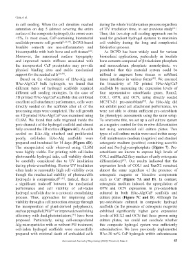
on 3D printed HAc-Alg/CaP was examined using for phenotypic assessments using the same setup.
CLSM. We found that cells migrated inside the To overcome this, we set up a cell culture system
pore channels of the hydrogel scaffold and almost as a negative control for this in vitro differentiation
fully covered the 3D surface (Figure 6C). As cells test using commercial cell culture plates. Two
seeded on HAc-Alg attached and proliferated types of cell culture media were used in this assay:
poorly, cell-laden HAc-Alg scaffolds were Cell maintenance medium (negative) and standard
prepared and incubated for 14 days (Figure 6D). osteogenic medium (positive) containing ascorbic
The encapsulated cells observed using CLSM acid and Na-β-glycerophosphate (Figure 7). Pre-
were highly viable. For printing with cell-laden osteoblasts are known to express high levels of
photocurable hydrogel inks, cell viability should COL1 and RunX2 (key markers of early osteogenic
be carefully considered due to UV irradiation differentiation) . Our results indicated that the
[42]
during photo-crosslinking. Shorter UV irradiation expression levels of COL1 and RunX2 remained
often leads to reasonably high cell viability even almost the same regardless of the presence of
though the mechanical stability of photocurable osteogenic reagents or bioactive components
hydrogels is compromised [21,41] . Indeed, there is such as CaP (Figure 7A and B). In contrast,
a significant trade-off between the mechanical osteogenic medium induced the upregulation of
performance and cell viability of cell-laden OPN and OCN expressions in pre-osteoblasts
hydrogel scaffolds due to a cytotoxic crosslinking cultured in both HAc-Alg/CaP scaffolds and
process. Thus, approaches for improving cell culture plates (Figure 7C and D). Although the
viability through a cell protection strategy through pre-osteoblasts cultured in composite hydrogel
the incorporation of plant-derived polyphenols, scaffolds (in the presence of osteogenic reagents)
such as pyrogallol (PG) or improved crosslinking exhibited significantly higher gene expression
[21]
efficiency with dual-photoinitiators, have been levels of RUX2 and OCN that those grown using
[41]
proposed. Particularly, using cell-encapsulated culture plates, we could not conclude whether
Alg microparticles with or without PG treatment, this composite hydrogel system was sufficiently
cell-laden hydrogel scaffolds were successfully osteoinductive. We have previously implemented
prepared with minimal death of embedded cells HAc-30 wt% CaP hydrogels within subcutaneous
International Journal of Bioprinting (2020)–Volume 6, Issue 2 43