Page 130 - IJB-10-6
P. 130

International Journal of Bioprinting                                 Fluid mechanics of extrusion bioprinting


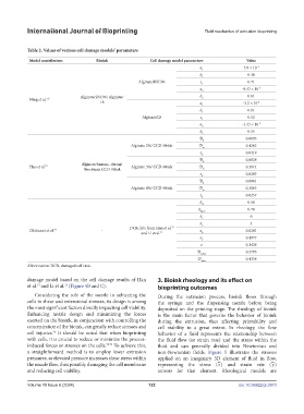
            Table 2. Values of various cell damage models’ parameters
             Model contributors        Bioink               Cell damage model parameters          Value
                                                                               a                3.9 × 10 -4
                                                                                1
                                                                               b                  0.48
                                                                                1
                                                       Alginate/RSC96          c                  0.71
                                                                                1
                                                                               a                -9.43 × 10 -4
                                                                                2
                                 Alginate/RSC96; Alginate/                     b 2                0.61
             Ning et al.  63
                                        L8                                     a 1              -3.2 × 10 -4
                                                                               b                  0.61
                                                                                1
                                                         Alginate/L8           c                  0.52
                                                                                1
                                                                               a                -1.43 × 10 -3
                                                                                2
                                                                               b 2                0.55
                                                                               D 0               0.0693
                                                    Alginate 2%/ CCD-986sk     D                 0.4262
                                                                                ∞
                                                                               ε                 0.0419
                                                                                d
                                                                               D 0               0.0828
                                 Alginate/human- dermal
             Han et al. 73                          Alginate 3%/ CCD-986sk     D                 0.3614
                                  fibroblasts CCD-986sk                         ∞
                                                                               ε d               0.0283
                                                                               D                 0.0861
                                                                                0
                                                    Alginate 4%/ CCD-986sk     D                 0.3063
                                                                                ∞
                                                                               ε                 0.0257
                                                                                d
                                                                               A                  0.50
                                                                                0
                                                                               A eq,∞             0.70
                                                                               k 1                 0
                                                                               k 2                 4
                                                    DCR data from Han et al.
                                                                    73
             Chirianni et al. 74         -                                     a                 0.0281
                                                        and Li et al. 75        p
                                                                               a                 0.4977
                                                                                e
                                                                                e                0.1428
                                                                              D e,max            0.2795
                                                                              D                  0.4358
                                                                                max
            Abbreviation: DCR, damaged cell ratio
            damage model based on the cell damage results of Han   3. Bioink rheology and its effect on
            et al.  and Li et al.  (Figure 4B and C).          bioprinting outcomes
                73
                          75
               Considering  the role  of  the  nozzle  in  subjecting  the   During the extrusion process, bioink flows through
            cells to shear and extensional stresses, its design is among   the syringe and the dispensing nozzle before being
            the most significant factors directly impacting cell viability.   deposited on the printing stage. The rheology of bioink
            Enhancing nozzle design and minimizing the forces   is the main factor that governs the behavior of bioink
            exerted on the bioink, in conjunction with controlling the   during the extrusion, thus affecting printability and
            concentration of the bioink, can greatly reduce stresses and   cell viability to a great extent. In rheology, the flow
            cell injuries.  It should be noted that when bioprinting   behavior of a fluid represents the relationship between
                      76
            with cells, it is crucial to reduce or minimize the process-  the fluid flow (or strain rate) and the stress within the
            induced forces or stresses on the cells. 73,74  To achieve this,   fluid and can generally divided into Newtonian  and
            a straightforward method is to employ lower extrusion   non-Newtonian fluids.  Figure  5 illustrates the stresses
            pressures, as elevated pressure increases shear stress within   applied on an imaginary 3D element of fluid in flow,
                                                                                                           =
                                                                                      =
            the nozzle flow, thus possibly damaging the cell membrane   representing the stress (τ ) and strain rate (γ )
            and reducing cell viability.                       tensors for that element. Rheological models are

            Volume 10 Issue 6 (2024)                       122                                doi: 10.36922/ijb.3973
   125   126   127   128   129   130   131   132   133   134   135