
Page 170 - IJB-10-6
P. 170
International Journal of Bioprinting 3D bioprinting technology for brain tumor
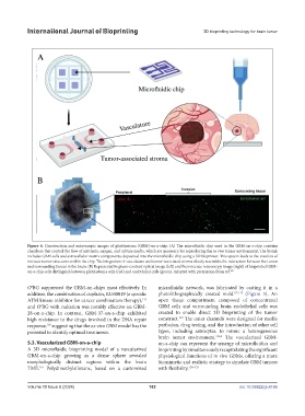
Figure 4. Construction and microscopic images of glioblastoma (GBM)-on-a-chip. (A) The microfluidic chip used in the GBM-on-a-chip contains
chambers that control the flow of nutrients, oxygen, and culture media, which are necessary for reproducing the in vivo tumor environment. The bioink
includes GBM cells and extracellular matrix components, deposited into the microfluidic chip using a 3D bioprinter. This system leads to the creation of
intricate tumor structures within the chip. The integration of vasculature and tumor-associated stroma closely resembles the interaction between the tumor
and surrounding tissues in the brain. (B) Representative phase-contrast optical image (left) and fluorescence microscopy image (right) of bioprinted GBM-
on-a-chip cells distinguish between glioblastoma cells (red) and endothelial cells (green). Adapted with permission from ref. 105
6
O BG suppressed the GBM-on-chips most effectively. In microfluidic network, was fabricated by casting it in a
addition, the combination of cisplatin, KU60019 (a specific photolithographically created mold 117,118 (Figure 5). An
ATM kinase inhibitor for cancer combination therapy), open tissue compartment composed of concentrated
115
and O BG with radiation was notably effective on GBM- GBM cells and surrounding brain endothelial cells was
6
28-on-a-chip. In contrast, GBM-37-on-a-chip exhibited created to enable direct 3D bioprinting of the tumor
119
high resistance to the drugs involved in the DNA repair construct. The outer channels were designed for media
response, suggesting that the ex vivo GBM model has the perfusion, drug testing, and the introduction of other cell
105
potential to identify optimal treatments. types, including astrocytes, to mimic a heterogeneous
brain tumor environment. 116,9 The vascularized GBM-
5.3. Vascularized GBM-on-a-chip on-a-chip can represent the synergy of microfluidics and
A 3D microfluidic bioprinting model of a vascularized bioprinting by simultaneously recapitulating the significant
GBM-on-a-chip growing as a dense sphere revealed physiological functions of in vivo GBMs, offering a more
morphologically distinct regions within the brain biomimetic and realistic strategy to simulate GBM tumors
TME. Polydimethylsiloxane, based on a customized with flexibility. 120–123
116
Volume 10 Issue 6 (2024) 162 doi: 10.36922/ijb.4166