
Page 171 - IJB-10-6
P. 171
International Journal of Bioprinting 3D bioprinting technology for brain tumor
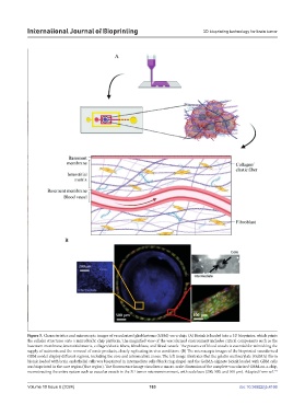
Figure 5. Characteristics and microscopic images of vascularized glioblastoma (GBM)-on-a-chip. (A) Bioink is loaded into a 3D bioprinter, which prints
the cellular structures onto a microfluidic chip platform. The magnified view of the vascularized environment includes critical components such as the
basement membrane, interstitial matrix, collagen/elastic fibers, fibroblasts, and blood vessels. The presence of blood vessels is essential for mimicking the
supply of nutrients and the removal of waste products, closely replicating in vivo conditions. (B) The microscopic images of the bioprinted vascularized
GBM model display different regions, including the core and intermediate zones. The left image illustrates that the gelatin methacrylate (GelMA) fibrin
bioink loaded with brain endothelial cells was bioprinted in intermediate cells (black ring shape) and the GelMA-alginate bioink loaded with GBM cells
was bioprinted in the core region (blue region). The fluorescence image visualizes a macro-scale dimension of the complete vascularized GBM-on-a-chip,
reconstructing the entire system such as vascular vessels in the 3D tumor microenvironment, with scale bars (200, 500, and 100 μm). Adapted from ref. 117
Volume 10 Issue 6 (2024) 163 doi: 10.36922/ijb.4166