
Page 166 - IJB-10-6
P. 166
International Journal of Bioprinting 3D bioprinting technology for brain tumor
vesicles and efflux transporters recruit astrocytes, the 3.1. Bioprinted TME models of brain tumors
invasion of GSCs and the secretion of anti-inflammatory 3D bioprinting technology has emerged as a potential tool
cytokines are promoted, creating an immunosuppressive for recapitulating the heterogeneous TME in various cancer
53
GBM microenvironment. Mesenchymal stem cells also types, including brain tumors. 3D-bioprinted brain tumor
play a major role in promoting mesenchymal-related models have been developed by optimizing the biomaterials
transcription and mediating tumor proliferation via and cell types according to their applications. As neural cell
9
interleukin-6 and exosome-encapsulating miRNA-1587. cultures require a complex microenvironment to retain the
54
These studies suggest that TME cells activated by cell-to- function of neurons and glial cells, biomaterials with good
cell interactions influence the development of treatment biocompatibility and mechanical properties are necessary
strategies for GBM. According to the type of cellular to support their function in 3D culture for brain tumor
components and applications, various printing methods research. For instance, while GelMA is used for culturing
3
are applied, including inkjet-based, extrusion-based, macrophages and astrocytes, which are essential cellular
and light-assisted bioprinting. Although inkjet-based components of GBM; microglia and human umbilical vein
55
bioprinting has a wide range of biological applications, it endothelial cells are cultured using collagen 10,11 (Figure 2).
is not appropriate for TME modeling due to limited cell Moreover, the critical properties of the cellular constituents
55
12
density. The 3D biomimetic models that reflect the TME vary depending on the biomaterials used. 3D co-culture
of GBM can be constructed by extrusion-based and DLP- systems using GelMA and GMHA include various
based 3D bioprinting. cellular compositions of GBM, such as astrocytes, GSCs,
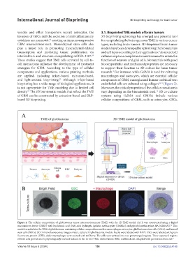
Figure 2. The cellular composition of glioblastoma tumor microenvironment (TME) with the 3D TME model. (A) It was constructed using a digital
56
micromirror device (DMD) with hyaluronic acid (HA)-rich hydrogels (gelatin methacrylate [GelMA] and glycidyl methacrylate-HA [GMHA]). This
model recapitulates the TME of glioblastoma, containing cellular compositions such as macrophages, astrocytes, glioblastoma stem cells (GSCs), and neural
stem cells (NSCs). (B) Immunofluorescence images of tetra-culture 3D glioblastoma models. Nuclei were labeled with DAPI. GSCs were labeled with green
fluorescent protein (GFP), while macrophages were stained with mCherry. The cells were printed into two prearranged regions. These separated regions
of both cells provide more physiologically relevant features to the in vivo TME. Abbreviation: RBC, red blood cell. Adapted with permission from ref. 56
Volume 10 Issue 6 (2024) 158 doi: 10.36922/ijb.4166