
Page 165 - IJB-10-6
P. 165
International Journal of Bioprinting 3D bioprinting technology for brain tumor
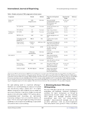
Table 1. Bioinks and printed TME components in brain tumors
Component Bioink Method Properties of 3D-printed Experimental Reference
model design
Glioma cell - GAF, Extrusion Expresses stability, In vitro and 29,30
(U118) transglutaminase upregulated EMT, stemness, in vivo
chemoresistance, and
tumorigenicity
GSC (GSC23) SA, gelatin Extrusion Exhibits proliferation and In vitro and 31,32
angiogenesis in vivo
GSC (GSC23) SA Extrusion Higher drug resistance In vitro 33,34
Glioblastoma GSC (SU3) GAF Extrusion Enhanced drug resistance In vitro 35,36
cell (U87) and cell viability
HUVECs, LFs GAF N/A Distinct angiogenesis and In vitro 37–38
EMT
GFP-expressing U87, MRCSs TPL Constructs the complex In vitro 40
hCMEC/D3 BBB system
Patient-derived BdECM or GBM-on-a-chip Incorporates heterogeneous In vitro 39,41
GBM, HUVECs collagen TME; demonstrates patient-
specific therapy resistance
- Matrigel MEW Facilitates cell growth, In vitro 42,43
migration, and drug
screening
Astrocytes Fibrinogen, Extrusion Feasible to test cell viability In vitro 4,44,45
alginate,genipin against drug
GSC (GSC23) - GAF Extrusion Alters cell morphology; In vitro 46,47
promotes vasculogenesis
and stemness
MSCs GAF Extrusion High cell viability; sufficient In vitro 47
cell–cell interaction
Macrophages, GMHA, GelMA DLP Upregulated GBM tissue- In vitro and 9,2
astrocytes, NPCs specific genes; activates in vivo
ECM and invasiveness
GASCs, microglia HA, RGD-alginate Extrusion Higher drug sensitivity In vitro 48,49
in TMZ and cisplatin;
maintains pluripotency
Abbreviations: BBB, blood–brain barrier; BdECM, brain decellularized extracellular matrix; DLP, digital light processing; ECM, extracellular matrix;
EMT, epithelial-mesenchymal transition; GAF, gelatin, alginate, fibrinogen; GASCs, glioma associated stromal cells; GBM, glioblastoma; GelMA, gelatin
methacrylate; GFP, green fluorescent protein; GMHA: glycidyl methacrylate-hyaluronic acid; GSC, glioblastoma stem cell; HA, hyaluronic acid; hCMEC/
D3, human cerebral microvascular endothelial cell line; HUVECs, human vascular endothelial cells; LFs, lung fibroblasts; MEW, melt electrowriting;
MRCSs, magnetically responsive cage-like scaffolds; MSCs, mesenchymal stem cells; NPCs, neural precursor cells; RGD-alginate, alginate modified by
Arg-Gly-Asp peptide sequence; SA, sodium alginate; TME, tumor microenvironment; TMZ, temozolomide; TPL, two-photon lithography.
the rapid printing speed is a significant advantage. 3. Mimicking the brain TME using
23
Similarly, laser-assisted printing can dispense formulated 3D bioprinting
cells drop-by-drop using a pulsed laser. It is highly
51
efficient compared to other methods because nozzles are The brain TME plays a pivotal role in tumor progression,
metabolism, and metastasis. Optimized physiological
unnecessary; therefore, it can deliver a higher viscosity and conditions for tumor development are formed by
density of cells with better viability. DLP and two-photon communication between various cell types, including
23
polymerization-based bioprinting are light-assisted GSCs, glioma cells, microglia, astrocytes, neurons,
bioprinting techniques that can be used for sensitive cell mesenchymal stem cells, and tumor-associated
types. Overall, selecting the right bioprinting method and microglia. Tumor-associated microglia with an M2
24
24
bioink has a critical impact on establishing an appropriate pro-tumor phenotype create an immunosuppressive
3D model for brain tumor modeling. TME and promote tumor growth.57 When extracellular
Volume 10 Issue 6 (2024) 157 doi: 10.36922/ijb.4166