
Page 196 - IJB-10-6
P. 196
International Journal of Bioprinting 3D-printed EVs for nasal septal defects
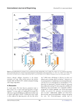
Figure 9. Immunohistochemical staining of Col II in rabbit nasal septal cartilage after 6 and 12 weeks (W) of repair. (A) Col II staining of the tissues.
Arrows denote deposited Col II. (B and C) Normalized analysis of Col II expression in the repaired tissues. **p < 0.01, ***p < 0.001, ****p < 0.0001. Scale
bars: 625 μm (A, black); 50 μm (A, white). Abbreviations: Ctrl: Control; Gel: Gelatin; PLGA: Polylactic acid-glycolic acid; EVs: Extracellular vesicles.
38
features, though collagen deposition in the tissues site. While stem cell therapy is effective, its safety and
increased over time. These results indicate that defects ethical aspects remain subjects of debate. Moreover, an
16
treated with Gel-PLGA+EVs exhibit enhanced healing increasing number of studies have demonstrated that stem
and collagen deposition. cells function in vivo through EVs as intermediaries. 39,40 In
this study, we developed a cell-free therapy for repairing
4. Discussion tissue defects by loading EVs into composite scaffolds and
In recent years, EVs have become prominent due to implanting them at the defect site. Composite scaffolds,
their crucial role in tissue repair and regulation of organ composed of GelMA hydrogel and PLGA-electrospun
13
function. In our study, we combined 3D-printed EVs biofilm, can provide the necessary microenvironment
41
with tissue-engineered biological scaffolds for the first time and structure. Additionally, GelMA and PLGA have
to repair nasal septum defects. Traditionally, tissue repair demonstrated good biocompatibility without damaging
is often achieved by injecting stem cells into the defect the cells. The PLGA-electrospun scaffold had an
Volume 10 Issue 6 (2024) 188 doi: 10.36922/ijb.4118