
Page 194 - IJB-10-6
P. 194
International Journal of Bioprinting 3D-printed EVs for nasal septal defects
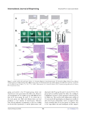
Figure 7. Animal models with nasal septal defects. (A) Schematic diagram of animal experiments. (B) Repaired cartilages obtained from different
treatment groups. (C) Defect healing rates at different time points. (D) Hematoxylin and eosin (H&E) staining at the defect site. Scale bars: 1 cm (B); 50
μm (D, white); 625 μm (D, black). **p < 0.01, ****p < 0.0001. Abbreviations: Ctrl: Control; W: Weeks; PLGA: Polylactic acid-glycolic acid; Gel: Gelatin;
EVs: Extracellular vesicles.
group, particularly in the EV-loaded group, where new observed in the EV group. By week 12, the Gel-PLGA+EVs
chondrocytes were generated at the defect site. However, group had successfully repaired the defect site, leading to
cell arrangement in the scaffold group still differed from a significant increase in newly generated chondrocytes at
that of the sham group. By week 12, the defect site in the defect site. The excellent anti-inflammatory ability of
the stent group was filled with fibrous tissue and had EVs prevented cell hypertrophy, resulting in an overall cell
produced a small number of chondrocytes. Toluidine and chondrocyte extracellular matrix arrangement that
blue staining indicated inflammation at this site, leading closely resembles that of the sham group. In contrast, due
to chondrocyte hypertrophy. A similar phenomenon was to the large defect size and inadequate cellular support
Volume 10 Issue 6 (2024) 186 doi: 10.36922/ijb.4118