
Page 195 - IJB-10-6
P. 195
International Journal of Bioprinting 3D-printed EVs for nasal septal defects
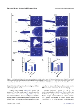
Figure 8. Toluidine blue staining of rabbit nasal septal cartilage after 6 and 12 weeks of repair. (A) Toluidine blue-stained tissues. (B and C) Normalized
analysis of the extent of cartilage matrix deposition in the repaired tissues at week 6 (B) and 12 (C). ***p < 0.001, ****p < 0.0001. Scale bars: 50 μm (A,
white); 625 μm (A, black). Abbreviations: Ctrl: Control; ns: Non-significant; Gel: Gelatin; PLGA: Polylactic acid-glycolic acid; EVs: Extracellular vesicles.
for proliferation and migration, the control group did not was observed in the scaffold group, there were noticeable
effectively repair the defect. differences when compared to the EV-treated group.
Toluidine blue staining (Figure 8A) indicates the Immunohistochemical staining of Col II was
presence of a cartilage matrix in newly formed tissue. The performed on the defect site (Figure 9) and revealed that
stained tissues were normalized (Figure 8B and C), and the EV-treated group at weeks 6 and 12 displayed more
the results revealed that the EV-treated group exhibited a Type 2 collagen expression than the control and Gel-
higher level of cartilage matrix deposition compared to the PLGA groups. Additionally, only some nascent mast
scaffold group. Although some cartilage matrix deposition chondrocytes in the Gel-PLGA group exhibited positive
Volume 10 Issue 6 (2024) 187 doi: 10.36922/ijb.4118