
Page 255 - IJB-10-6
P. 255
International Journal of Bioprinting 3D-printed contractive pennate muscle
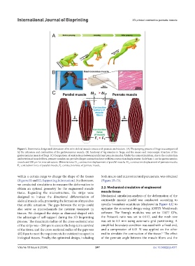
Figure 1. Biomimetic design and fabrication of in vitro skeletal muscle tissues with pennate architecture. (A) The jumping process of frogs is accompanied
by the extension and contraction of the gastrocnemius muscle. (B) Anatomy of leg muscles in frogs, and the macro and microscopic structure of the
gastrocnemius muscle of frogs. (C) Comparison of contractions between parallel and pennate muscles. Under the same stimulation, due to the contraction
and rotation of muscle fibers, pennate muscles can provide a larger contraction force with less contraction displacement. Scale bars: 1 cm for gastrocnemius
muscle and 200 μm for microstructure. Abbreviations: ΔL : contraction displacement of parallel muscle; ΔL : contraction displacement of pennate muscle;
P
L
F : contraction force of parallel muscle; F : contraction force of pennate muscle.
L P
within a certain range to change the shape of the tissues both macro and microstructural parameters, was obtained
(Figures S1 and S2, Supporting Information). Furthermore, (Figure 3B–D).
we conducted simulations to measure the deformation to
obtain an optimal geometry for the engineered muscle 2.2. Mechanical simulation of engineered
tissue. Regarding the microstructure, the strips were muscle tissue
designed to induce the directional differentiation of Mechanical simulation analysis of the deformation of the
skeletal muscle cells, promoting the formation of myotubes contractile muscle model was conducted according to
that enable actuation. The gaps between the strips could specific boundary conditions (displayed in Figure 4A) to
also serve as microchannels for nutrient transport in optimize the structural design using ANSYS Workbench
tissues. We designed the strips as diamond-shaped with software. The Young’s modulus was set to 12077 GPa,
the advantage of self-support during the 3D bioprinting the Poisson’s ratio was set to 0.117, and the mesh size
process. The maximum radius of the cross-sectional area was set to 0.1 mm using automatic grid partitioning. A
of the strips was ~200 μm to ensure the long-term survival simplified boundary condition was used with a fixed end,
of the tissue, and the cross-sectional radius of the gaps was and a compression of 0.01 N was applied on the other
21
482.8 μm to meet the requirements for nutrient transport in end to simulate the contraction of the tissue. The effect
biological tissues. Finally, the optimized design, including of the pennate angle between the muscle fibers and the
Volume 10 Issue 6 (2024) 247 doi: 10.36922/ijb.4371