
Page 258 - IJB-10-6
P. 258
International Journal of Bioprinting 3D-printed contractive pennate muscle
cell viability assessment. Immunol staining fix solution for reliable printability and shape fidelity. Here, bioink
(P0098; Beyotime, China), Triton X-100 (T8200; consisting of GelMA (at a 60% degree of substitution) and
Solarbio, China), Alexa Fluor 488-conjugated phalloidin C2C12 myoblasts (under five passages) was selected for 3D
(A12379; Invitrogen, USA) (affinity [Kd]: 20 nM), and bioprinting. The GelMA was dissolved at a concentration
4ʹ,6-diamidino-2-phenylindole staining solution (DAPI; of 5% in PBS with 0.25% w/v LAP, and heated in a water
C1006; Beyotime, China) were utilized for fluorescence bath at 60°C for 30 min. The mixture was then refrigerated
staining. Polydimethylsiloxane (PDMS; Sylgard 184; Dow at 4°C for 30 min and thawed at 37°C. The C2C12 cells
Corning, USA) was used to fabricate U-shape posts. were cultured in GM at 37°C and 5% CO atmosphere
2
and passaged at 80% confluency. For the 3D bioprinting
2.4. 3D printing and cultivation of skeletal experiments, the cells were digested with 0.25% trypsin-
muscle tissues EDTA at about 80% confluency and mixed with GelMA
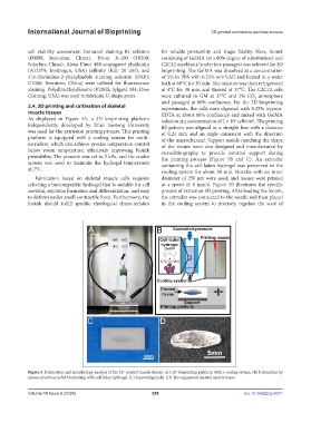
As displayed in Figure 5A, a 3D bioprinting platform solution at a concentration of 2 × 10 cells/mL. The printing
6
independently developed by Xi’an Jiaotong University fill pattern was aligned in a straight line with a distance
was used for the extrusion printing process. This printing of 0.23 mm and an angle consistent with the direction
platform is equipped with a cooling system for multi- of the microchannel. Support molds matching the shape
extruders, which can achieve precise temperature control of the tissues were also designed and manufactured by
below room temperature, effectively improving bioink stereolithography to provide external support during
printability. The pressure was set to 3 kPa, and the cooler the printing process (Figure 5B and C). An extruder
system was used to maintain the hydrogel temperature containing the cell-laden hydrogel was preserved in the
at 2°C. cooling system for about 30 min. Nozzles with an inner
Fabrication based on skeletal muscle cells requires diameter of 250 μm were used, and tissues were printed
selecting a biocompatible hydrogel that is suitable for cell at a speed of 8 mm/s. Figure 5B illustrates the specific
survival, myotube formation and differentiation, and easy process of extrusion 3D printing. After loading the bioink,
to deform under small contractile force. Furthermore, the the extruder was connected to the nozzle and then placed
bioink should fulfill specific rheological characteristics in the cooling system to precisely regulate the state of
Figure 5. Fabrication and morphology analysis of the 3D-printed muscle tissues. (A) 3D-bioprinting platform with a cooling system. (B) Fabrication by
means of extrusion 3D bioprinting with cell-laden hydrogel. (C) Supporting mold. (D) The engineered skeletal muscle tissue.
Volume 10 Issue 6 (2024) 250 doi: 10.36922/ijb.4371