
Page 263 - IJB-10-6
P. 263
International Journal of Bioprinting 3D-printed contractive pennate muscle
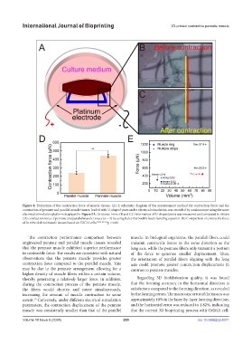
Figure 8. Evaluation of the contraction force of muscle tissues. (A) A schematic diagram of the measurement method for contraction force and the
contraction of pennate and parallel muscle tissues, loaded with U-shaped posts under electrical stimulation, was recorded by a microscope using the same
electrical stimulation platform displayed in Figure 5A. Grid size: 1mm. (B and C) Deformation of U-shaped posts was measured and computed to obtain
(D) contraction force of pennate and parallel muscle tissues (n = 3) by using Euler-Bernoulli’s beam bending equation. (E) Comparison of contractile force
of in vitro skeletal muscle tissues based on C2C12 cells. 20,36–40 **p < 0.01.
The contraction performance comparison between muscle. In biological organisms, the parallel fibers could
engineered pennate and parallel muscle tissues revealed transmit contractile forces in the same direction as the
that the pennate muscle exhibited superior performance long axis, while the pennate fibers only transmit a portion
in contractile force. The results are consistent with natural of the force to generate smaller displacement. Thus,
observations that the pennate muscle provides greater the orientation of parallel fibers aligning with the long
contraction force compared to the parallel muscle. This axis could promote greater contraction displacement in
may be due to the pennate arrangement allowing for a contrast to pennate muscles.
higher density of muscle fibers within a certain volume,
thereby generating a relatively larger force. In addition, Regarding 3D biofabrication quality, it was found
during the contraction process of the pennate muscle, that the forming accuracy in the horizontal direction is
the fibers would shorten and rotate simultaneously, satisfactory compared to the forming direction, as revealed
increasing the amount of muscle contraction to some by the forming errors. The macro size error of the tissues was
extent. Conversely, under different electrical stimulation approximately 10% in the layer-by-layer forming direction,
30
parameters, the contraction displacement of the pennate and the horizontal error was reduced to 1.82%, indicating
muscle was consistently smaller than that of the parallel that the current 3D bioprinting process with GelMA cell-
Volume 10 Issue 6 (2024) 255 doi: 10.36922/ijb.4371