
Page 335 - IJB-10-6
P. 335
International Journal of Bioprinting 3D-printed PPDO/GO stents for CHD treatment
Vascular stents are one of the medical devices With the incorporation of GO, the quantity of adhered
that are directly in contact with blood; hence, the platelets decreases. According to Figure 6c, the number
hemocompatibility of PPDO/GO stents needs to be of adhered platelets per unit area for PPDO/0.2%GO
evaluated to avoid hemolysis and thrombosis. During and PPDO/0.5%GO are 6853.25 ± 1237.32 and 4405.66
−2
hemolysis, RBCs rupture; free hemoglobin, intracellular ± 1207.06 mm , respectively, and are significantly lower
−2
components, and pro-thrombotic substances are released than that of pristine PPDO (12346.72 ± 1225.30 mm ).
into the plasma, activating the coagulation cascade and Additionally, there is a significant statistical difference
leading to thrombosis. According to the ISO 10993-4 between the PPDO/0.2% and PPDO/0.5% groups. The
92
standard, a hemolysis rate lower than 5% is acceptable for SEM images (Figure 6d–f) also demonstrate the trend of
blood-contacting biomaterials. Results of the hemolysis a smaller quantity of adhered platelets with the increase of
test (Figure 6a and b) revealed that the hemolysis rates of GO content. The adhered platelets in all groups demonstrate
all samples are below 1%, indicating that PPDO/GO stents a round shape without protruded pseudopodia, suggesting
are acceptable and do not cause hemolysis. a non-activated state. GO introduces a large number of
hydrophilic groups, which conduce albumin adsorption
After adherence to the stent surface, platelets can be and competitively inhibit fibrinogen adsorption. This
activated and aggregate, causing thrombosis, inflammation, forms a protective layer on the surface of the material to
and even restenosis. 93,94 Therefore, it is essential to evaluate prevent the direct contact of platelets, thus reducing the
the level of platelet adhesion of PPDO/GO stents. adhesion and activation of platelets. 95
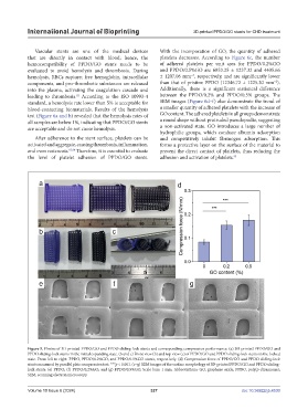
Figure 5. Photos of 3D-printed PPDO/GO and PPDO sliding-lock stents and corresponding compression performance. (a) 3D-printed PPDO/GO and
PPDO sliding-lock stents in the initial expanding state. (b and c) Front view (b) and top view (c) of PPDO/GO and PPDO sliding-lock stents in the locked
state. From left to right: PPDO, PPDO/0.2%GO, and PPDO/0.5%GO stents, respectively. (d) Compression force of PPDO/GO and PPDO sliding-lock
stents measured by parallel plate compression test. ***p < 0.001. (e–g) SEM images of the surface morphology of 3D-printed PPDO/GO and PPDO sliding-
lock stents: (e) PPDO; (f) PPDO/0.2%GO; and (g) PPDO/0.5%GO. Scale bars: 1 mm. Abbreviations: GO, graphene oxide; PPDO, poly(p-dioxanone);
SEM, scanning electron microscopy.
Volume 10 Issue 6 (2024) 327 doi: 10.36922/ijb.4530