
Page 336 - IJB-10-6
P. 336
International Journal of Bioprinting 3D-printed PPDO/GO stents for CHD treatment.
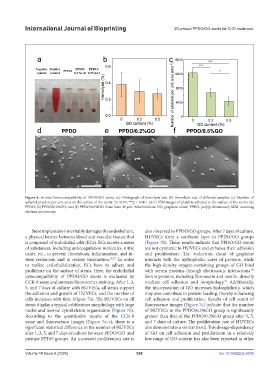
Figure 6. In vitro hemocompatibility of PPDO/GO stents. (a) Photograph of hemolysis test. (b) Hemolysis rate of different samples. (c) Number of
adhered platelets per unit area on the surface of the stents. *p <0.05, ***p < 0.001. (d–f) SEM images of platelets adhered to the surface of the stents: (d)
PPDO; (e) PPDO/0.2%GO; and (f) PPDO/0.5%GO. Scale bars: 20 μm. Abbreviations: GO, graphene oxide; PPDO, poly(p-dioxanone); SEM, scanning
electron microscopy.
Stent implantation inevitably damages the endothelium, also observed in PPDO/GO groups. After 7 days of culture,
a physical barrier between blood and vascular tissues that HUVECs form a confluent layer in PPDO/GO groups
is composed of endothelial cells (ECs). ECs secrete a series (Figure 7b). These results indicate that PPDO/GO stents
of substances, including anticoagulation molecules, nitric are not cytotoxic to HUVECs and enhance their adhesion
oxide, etc., to prevent thrombosis, inflammation, and in- and proliferation. The π-electron cloud of graphene
stent restenosis, and to restore vasomotion. 96,97 In order interacts with the hydrophobic cores of proteins, while
to realize endothelialization, ECs have to adhere and the high-density oxygen-containing groups of GO bind
proliferate on the surface of stents. Here, the endothelial with serum proteins through electrostatic interactions.
98
cytocompatibility of PPDO/GO stents is evaluated by Serum proteins, including fibronectin and insulin, directly
CCK-8 assay and immunofluorescence staining. After 1, 3, mediate cell adhesion and morphology. Additionally,
99
5, and 7 days of culture with HUVECs, all stents support the incorporation of GO increases hydrophilicity, which
the adhesion and growth of HUVECs, and the number of may also contribute to protein loading, thereby enhancing
cells increases with time (Figure 7a). The HUVECs on all cell adhesion and proliferation. Results of cell count of
stents display a typical cobblestone morphology with large fluorescence images (Figure 7c) indicate that the number
nuclei and normal cytoskeleton organization (Figure 7b). of HUVECs in the PPDO/0.5%GO group is significantly
According to the quantitative results of the CCK-8 greater than that of the PPDO/0.2%GO group after 3, 5,
assay and fluorescence images (Figure 7a–c), there is a and 7 days of culture. The proliferation rate of HUVECs
significant statistical difference in the number of HUVECs also demonstrates a similar trend. This dosage-dependency
after 1, 3, 5, and 7 days of culture between PPDO/GO and of GO on cell adhesion and proliferation in a relatively
pristine PPDO groups. An increased proliferation rate is low range of GO content has also been reported in other
Volume 10 Issue 6 (2024) 328 doi: 10.36922/ijb.4530