
Page 338 - IJB-10-6
P. 338
International Journal of Bioprinting 3D-printed PPDO/GO stents for CHD treatment.
studies, 51,100 one of which discovered that 0.5% GO- significant in the PPDO/0.5%GO group, similar to that
incorporated thermoplastic polyurethane presented higher of normal endothelium. The H&E staining (Figure 9a–c)
HUVEC attachment and proliferation. 100 revealed that, after implantation in the abdominal aortas
of SD rats for 4 weeks, intimal hyperplasia was observed
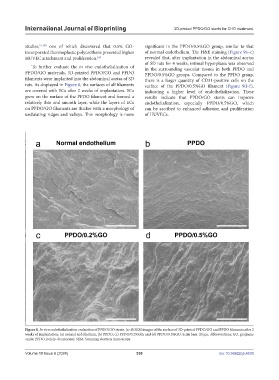
To further evaluate the in vivo endothelialization of in the surrounding vascular tissues in both PPDO and
PPDO/GO materials, 3D-printed PPDO/GO and PPDO PPDO/0.5%GO groups. Compared to the PPDO group,
filaments were implanted into the abdominal aortas of SD there is a larger quantity of CD31-positive cells on the
rats. As displayed in Figure 8, the surfaces of all filaments surface of the PPDO/0.5%GO filament (Figure 9d–f),
are covered with ECs after 2 weeks of implantation. ECs indicating a higher level of endothelialization. These
grew on the surface of the PPDO filament and formed a results indicate that PPDO/GO stents can improve
relatively thin and smooth layer, while the layers of ECs endothelialization, especially PPDO/0.5%GO, which
on PPDO/GO filaments are thicker with a morphology of can be ascribed to enhanced adhesion and proliferation
undulating ridges and valleys. This morphology is more of HUVECs.
Figure 8. In vivo endothelialization evaluation of PPDO/GO stents. (a–d) SEM images of the surface of 3D-printed PPDO/GO and PPDO filaments after 2
weeks of implantation: (a) normal endothelium; (b) PPDO; (c) PPDO/0.2%GO; and (d) PPDO/0.5%GO. Scale bars: 20 μm. Abbreviations: GO, graphene
oxide; PPDO, poly(p-dioxanone); SEM, Scanning electron microscopy.
Volume 10 Issue 6 (2024) 330 doi: 10.36922/ijb.4530