
Page 469 - IJB-10-6
P. 469
International Journal of Bioprinting Stress prediction in 3D-printed scaffolds
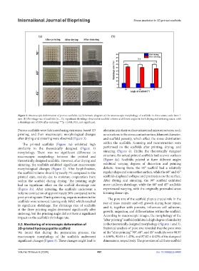
Figure 3. Macroscopic deformation of porous scaffolds. (a) Schematic diagram of the macroscopic morphology of scaffolds in three states; scale bars: 1
mm. (b) Shrinkage rate of scaffolds (n = 3); significant shrinkage observed in scaffold volume at different angles in both drying and sintering states, with
a shrinkage rate of 30% after sintering. ***p < 0.001; N.S., not significant.
Porous scaffolds were fabricated using extrusion-based 3D alterations in their surface or internal microstructures, such
printing, and their macroscopic morphological changes as variations in the stress contact surface, filament diameter,
after drying and sintering were observed (Figure 3). and scaffold porosity, which affect the stress distribution
The printed scaffolds (Figure 3a) exhibited high within the scaffolds. Scanning and reconstruction were
similarity to the theoretically designed (Figure 1) performed on the scaffolds after printing, drying, and
morphology. There was no significant difference in sintering (Figure 4). Unlike the theoretically designed
macroscopic morphology between the printed and structure, the actual printed scaffolds had uneven surfaces
theoretically designed scaffolds. However, after drying and (Figure 4a). Scaffolds printed at three different angles
sintering, the scaffolds exhibited significant macroscopic exhibited varying degrees of distortion and printing
morphological changes (Figure 3). After lyophilization, defects. Among them, the 90° scaffold had a relatively
the scaffold volume shrank by nearly 7% compared to the regular shape and a smoother surface, while the 60° and 45°
printed state, mainly due to moisture evaporation from scaffolds displayed collapse and protrusion on the surface.
within the scaffold during drying. The printing angle After drying and sintering, the 90° scaffold exhibited
had no significant effect on the scaffold shrinkage rate more uniform shrinkage, while the 60° and 45° scaffolds
(Figure 3b). After sintering, the scaffolds underwent a experienced warping, with the originally protruded areas
volume contraction of approximately 30% compared to the forming dense tips.
post-printing state. During sintering, organic matters in the The pore size of the scaffold plays a crucial role in the
scaffolds were removed, leaving only HAP, which resulted rate of mass transfer and cell growth during bone repair,
in significant shrinkage. The shrinkage rate of scaffolds and it, together with porosity, influences cell adhesion,
at the three printing angles decreased significantly after growth, migration, and differentiation within the scaffold.
sintering, but the printing angle did not have a significant According to macroscopic images, the morphology of the
impact on the scaffold’s shrinkage rate.
“after printing” scaffold exhibited a high degree of similarity
3.2. Monitoring of microscopic deformation of to the theoretically designed morphology (Figures 1 and 3).
3D-printed hydroxyapatite scaffold Statistical analysis of pore size revealed that the pore sizes
We found that during the preparation process, the of the “after printing” 90°, 60°, and 45° scaffolds were 90.97
macroscopic morphology of the scaffolds underwent ± 4.00%, 90.63 ± 1.45%, and 87.02 ± 4.42% of the designed
significant changes (Figure 3). These changes might lead to dimensions, respectively. The pore sizes of all three scaffold
Volume 10 Issue 6 (2024) 461 doi: 10.36922/ijb.4460