
Page 470 - IJB-10-6
P. 470
International Journal of Bioprinting Stress prediction in 3D-printed scaffolds
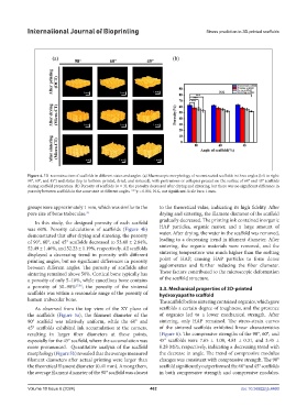
Figure 4. 3D reconstruction of scaffolds in different states and angles. (a) Macroscopic morphology of reconstructed scaffolds in three angles (left to right:
90°, 60°, and 45°) and states (top to bottom: printed, dried, and sintered), with protrusions or collapses present on the surface of 60° and 45° scaffolds
during scaffold preparation. (b) Porosity of scaffolds (n = 3); the porosity decreased after drying and sintering, but there was no significant difference in
porosity between scaffolds in the same state at different angles. ***p < 0.001; N.S., not significant. Scale bars: 1 mm.
groups were approximately 1 mm, which was similar to the to the theoretical value, indicating its high fidelity. After
pore size of bone trabeculae. drying and sintering, the filament diameter of the scaffold
35
gradually decreased. The printing ink contained inorganic
In this study, the designed porosity of each scaffold
was 60%. Porosity calculations of scaffolds (Figure 4b) HAP particles, organic matter, and a large amount of
demonstrated that after drying and sintering, the porosity water. After drying, the water in the scaffold was removed,
of 90°, 60°, and 45° scaffolds decreased to 53.48 ± 2.64%, leading to a decreasing trend in filament diameter. After
52.49 ± 1.46%, and 52.23 ± 1.19%, respectively. All scaffolds sintering, the organic materials were removed, and the
displayed a decreasing trend in porosity with different sintering temperature was much higher than the melting
printing angles, but no significant differences in porosity point of HAP, causing HAP particles to form dense
between different angles. The porosity of scaffolds after agglomerates and further reducing the fiber diameter.
sintering remained above 50%. Cortical bone typically has These factors contributed to the microscopic deformation
a porosity of only 5–10%, while cancellous bone contains of the scaffold structure.
a porosity of 50–90% 27,36 ; the porosity of the sintered 3.3. Mechanical properties of 3D-printed
scaffolds was within a reasonable range of the porosity of hydroxyapatite scaffold
human trabecular bone. The scaffold before sintering contained organics, which gave
As observed from the top view of the XY plane of scaffolds a certain degree of toughness, and the presence
the scaffolds (Figure 5a), the filament diameter of the of organics led to a lower mechanical strength. After
90° scaffold was relatively uniform, while the 60° and sintering, only HAP remained. The stress-strain curves
45° scaffolds exhibited ink accumulation at the corners, of the sintered scaffolds exhibited linear characteristics
resulting in larger fiber diameters at these points, (Figure 6). The compressive strengths of the 90°, 60°, and
especially for the 45° scaffold, where the accumulation was 45° scaffolds were 7.65 ± 1.08, 4.81 ± 0.31, and 3.45 ±
more pronounced. Quantitative analysis of the scaffold 0.28 MPa, respectively, indicating a decreasing trend with
morphology (Figure 5b) revealed that the average measured the decrease in angle. The trend of compressive modulus
filament diameters after actual printing were larger than changes was consistent with compressive strength. The 90°
the theoretical filament diameter (0.41 mm). Among them, scaffold significantly outperformed the 60° and 45° scaffolds
the average filament diameter of the 90° scaffold was closest in both compressive strength and compressive modulus.
Volume 10 Issue 6 (2024) 462 doi: 10.36922/ijb.4460