
Page 100 - IJB-7-2
P. 100
3D Printed and Ion Controllable Release
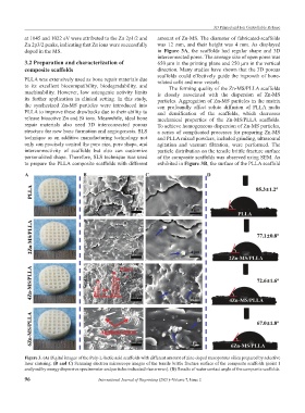
at 1045 and 1022 eV were attributed to the Zn 2p1/2 and amount of Zn-MS. The diameter of fabricated-scaffolds
Zn 2p3/2 peaks, indicating that Zn ions were successfully was 12 mm, and their height was 4 mm. As displayed
doped in the MS. in Figure 3A, the scaffolds had regular shape and 3D
interconnected pores. The average size of open pores was
3.2 Preparation and characterization of 650 μm in the printing plane and 250 μm in the vertical
composite scaffolds direction. Many studies have shown that the 3D porous
scaffolds could effectively guide the ingrowth of bone-
PLLA was extensively used as bone repair materials due related cells and new vessels.
to its excellent biocompatibility, biodegradability, and The forming quality of the Zn-MS/PLLA scaffolds
machinability. However, low osteogenic activity limits is closely associated with the dispersion of Zn-MS
its further application in clinical setting. In this study, particles. Aggregation of Zn-MS particles in the matrix
the synthesized Zn-MS particles were introduced into can profoundly affect solute diffusion of PLLA melts
PLLA to improve these drawbacks due to their ability to and densification of the scaffolds, which decreases
release bioactive Zn and Si ions. Meanwhile, ideal bone mechanical properties of the Zn-MS/PLLA scaffolds.
repair materials also need 3D interconnected porous To achieve homogeneous dispersion of Zn-MS particles,
structure for new bone formation and angiogenesis. SLS a series of complicated processes for preparing Zn-MS
technique as an additive manufacturing technology not and PLLA mixed powders, included grinding, ultrasound
only can precisely control the pore size, pore shape, and agitation and vacuum filtration, were performed. The
interconnectivity of scaffolds but also can customize particle distribution on the tensile brittle fracture surface
personalized shape. Therefore, SLS technique was used of the composite scaffolds was observed using SEM. As
to prepare the PLLA composite scaffolds with different exhibited in Figure 3B, the surface of the PLLA scaffold
A B C D
Figure 3. (A) Digital images of the Poly-L-lactic acid scaffolds with different amount of zinc-doped mesoporous silica prepared by selective
laser sintering. (B and C) Scanning electron microscope images of the tensile brittle fracture surface of the composite scaffolds (point 1
analyzed by energy dispersive spectrometer and particles indicated blue arrows). (D) Results of water contact angle of the composite scaffolds.
96 International Journal of Bioprinting (2021)–Volume 7, Issue 2