
Page 103 - IJB-7-2
P. 103
Qian, et al.
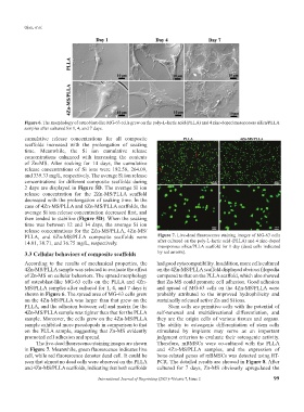
Figure 6. The morphology of osteoblast-like MG-63 cells grew on the poly-L-lactic acid (PLLA) and 4 zinc-doped mesoporous silica/PLLA
samples after cultured for 1, 4, and 7 days.
cumulative release concentrations for all composite
scaffolds increased with the prolongation of soaking
time. Meanwhile, the Si ion cumulative release
concentrations enhanced with increasing the contents
of Zn-MS. After soaking for 14 days, the cumulative
release concentrations of Si ions were 182.58, 264.09,
and 339.33 mg/L, respectively. The average Si ion release
concentrations for different composite scaffolds during
2 days are displayed in Figure 5D. The average Si ion
release concentration for the 2Zn-MS/PLLA scaffold
decreased with the prolongation of soaking time. In the
case of 4Zn-MS/PLLA and 6Zn-MS/PLLA scaffolds, the
average Si ion release concentration decreased first, and
then tended to stabilize (Figure 5D). When the soaking
time was between 12 and 14 days, the average Si ion
release concentrations for the 2Zn-MS/PLLA, 4Zn-MS/
PLLA, and 6Zn-MS/PLLA composite scaffolds were Figure 7. Live-dead fluorescence staining images of MG-63 cells
14.01, 30.71, and 36.75 mg/L, respectively. after cultured on the poly-L-lactic acid (PLLA) and 4 zinc-doped
mesoporous silica/PLLA scaffold for 1 day (dead cells indicated
3.3 Cellular behaviors of composite scaffolds by red arrows).
According to the results of mechanical properties, the had good cytocompatibility. In addition, more cells cultured
4Zn-MS/PLLA sample was selected to evaluate the effect on the 4Zn-MS/PLLA scaffold displayed obvious filopodia
of Zn-MS on cellular behaviors. The spread morphology compared to that on the PLLA scaffold, which also showed
of osteoblast-like MG-63 cells on the PLLA and 4Zn- that Zn-MS could promote cell adhesion. Good adhesion
MS/PLLA samples after cultured for 1, 4, and 7 days is and spread of MG-63 cells on the 4Zn-MS/PLLA were
shown in Figure 6. The spread area of MG-63 cells grew probably attributed to the improved hydrophilicity and
on the 4Zn-MS/PLLA was larger than that grew on the sustainedly released active Zn and Si ions.
PLLA, and the adhesion between cell and matrix for the Stem cells are primitive cells with the potential of
4Zn-MS/PLLA sample was tighter than that for the PLLA self-renewal and multidirectional differentiation, and
sample. Moreover, the cells grew on the 4Zn-MS/PLLA they are the origin cells of various tissues and organs.
sample exhibited more pseudopods in comparison to that The ability to osteogenic differentiation of stem cells
on the PLLA sample, suggesting that Zn-MS evidently stimulated by implants may serve as an important
promoted cell adhesion and spread. judgment criterion to evaluate their osteogenic activity.
The live-dead fluorescence staining images are shown Therefore, mBMSCs were co-cultured with the PLLA
in Figure 7. Meanwhile, green fluorescence indicates live and 4Zn-MS/PLLA samples, and the expression of
cell, while red fluorescence denotes dead cell. It could be bone-related genes of mBMSCs was detected using RT-
seen that almost no dead cells were observed on the PLLA PCR. The detailed results are showed in Figure 8. After
and 4Zn-MS/PLLA scaffolds, indicating that both scaffolds cultured for 7 days, Zn-MS obviously upregulated the
International Journal of Bioprinting (2021)–Volume 7, Issue 2 99