
Page 73 - IJB-7-2
P. 73
González, et al.
defining element that differentiates “bioprinting” from articles were examined to find the trends in the intended
“3D printing.” The success of generating functional applications, the bioprinting technologies used and the
tissues relies heavily on the quality of the bioink. design/composition of the bioinks employed (cell types,
Research groups around the globe have devoted hydrogels, and functional additives).
their efforts to developing protocols and bioink-related
technologies that are bringing us closer to the ambitious 3. Applications
goal of bioprinting fully functional tissues and organs. Figure 2 shows the most reported applications revealed
In this work, we describe the present landscape in the final pool of selected articles. The outcome of
of bioink use and development, as reported in 393 our analysis reveals the two main reasons that motivate
original research papers published from January 2000 to the research and use of bioinks: (i) The development of
June 2019. We also discuss the trends revealed by this bioprinting technology (generic) and (ii) clinical needs.
scientometric analysis from a technical perspective. We Approximately one third of the analyzed papers focused
start by presenting and discussing the most frequently on the development of new materials to formulate bioinks
reported applications in bioprinting and the most or the introduction of novel bioprinting strategies. This
commonly used bioprinting techniques. We then describe observation was expected as bioprinting is an innovative
the trends related to the three main components of the technology currently transitioning through an early
bioinks: Cells, hydrogels, and functional additives. development stage.
2. Information search methodology The development of bioprinting strategies [11,12] and
bioprinters with novel features has become a frequent
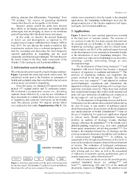
We conducted document search using the Scopus database. endeavor of engineers in academia and industry . The
[13]
Figure 1 presents the terms and search criteria used. We portfolio of bioprinting methods and bioprinters has
considered words used in the literature as synonyms of greatly evolved in the last two decades. The original
bioink and excluded terms that could lead to the inclusion devices were less complex [14-16] and adapted to perform
of documents not related to bioprinting. proof-of-principle experiments and demonstrate the
From this query, we obtained 529 manuscripts that deposition of drops containing living cells in a single layer
include 457 original articles and 72 conference papers. (and their short-term survival). These have now evolved
We conducted a de-duplication process (i.e., discarding into sophisticated designs that enable multi-material and
replicate items) followed by a one-by-one validation of multi-cell type fabrication of multi-layered constructs in
these documents to confirm that all of them were related the size range of ~ cm at resolutions of ~ 10 µm .
[17]
3
to bioprinting and contained information on the bioinks The development of bioprinting devices and bioink
used. This process yielded 393 original articles which formulations has also advanced in a parallel fashion during
were analyzed in this study (Supplementary File 1). The the past 20 years. A vast number of published papers
about bioprinting have focused more on the development
and characterization of bioink formulations [18,19] than on
the use of bioprinting for a particular application aligned
to clinical needs. Bioink characterization frequently
involves an analysis of rheology, because rheology
strongly influences the printability of the bioink under
[20]
different printing conditions (e.g., flow rates, printhead
linear speed, temperature, and printing pressure) [18,21] . For
instance, the interplay between the ink rheology and the
printing parameters determines the fidelity and resolution
of extrusion-based bioprinting [12,20,21] . In addition, an
assessment of the biological performance of the bioink is
practically mandatory in papers related to the development
of bioinks. The evaluation of biological performance
includes the determination of cell viability immediately
after printing and over time, as well as the assessment
of indicators of metabolic activity, proliferation, gene or
protein expression, and/or differentiation .
[18]
When it comes to clinical needs, the most frequently
Figure 1. Schematic representation of the query terminology used reported bioprinted tissues are cartilage [22-24] and
for the data search. The asterisks (*) mark root words and indicate bone [25-27] , representing 16% and 11% of the analyzed
that all possible suffixes were covered in the query. documents, respectively (Figure 2). The statistics reveal
International Journal of Bioprinting (2021)–Volume 7, Issue 2 69