
Page 78 - IJB-7-2
P. 78
A Scientometric Analysis
consideration on viscosity , cell density, and interaction technologies aimed at generating a safe and effective
[99]
with the receiving substrate (i.e., the surface tension and cell source for clinical use [112-114] . Nevertheless, the vast
wettability). majority of the bioprinting studies conducted today
Extrusion bioprinting requires several other use cells to develop proof-of-concept tissue constructs
innovations. Many of these are related to the ability to rather than functional tissues for transplantation [111,115] .
co-extrude two or more different materials concurrently As a general rule, the bioink design and the
(multimaterial bioprinting) [78,100-102] . For instance, these bioprinting conditions for tissue constructs, whether
materials may contain different types of cells or different for transplantation or for ex-vivo applications,
hydrogels to recapitulate the composition and architecture should favor cell viability for extended periods, cell
of a native tissue. In this context, a significant challenge proliferation, and the capability to develop into mature
is the selection/design of a compatible set of bioinks in tissues (assessed, e.g., by protein expression and
terms of rheology, interfacial tension, and co-flowability, immunohistochemistry) [116-118] . Within this framework,
among other properties [103] . cells can be purchased (i.e., from the American Type
Novel embodiments of extrusion bioprinting have Culture Collection or other cell culture companies [56,119]
greatly pushed the limits of biofabrication in the last or harvested from primary tissues [120,121] ).
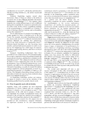
5 years. For example, innovative bioprinting heads that Figure 6 presents the most reported cell types used in
use chaotic static mixers [102] to fabricate microstructured bioink formulations. At first sight, no direct correlation is
hydrogel filaments are enabling new applications, observed between the applications (Figure 2) and the cells
such as the facile fabrication of muscle-like fibers . used (Figure 6). Notably, the utilization of cells usually
[12]
Extrusion-based bioprinters are also becoming more serves one of two purposes: Biofabrication of a specific
portable, and hand-held models promise to enable in situ tissue or organ, or assessment of the performance of a
and in vivo bioprinting for a wide range of scenarios, bioink material or a bioprinting technology (technology
including wound healing, dentistry, and minor and major development). Stem cells (SCs) and induced pluripotent
surgeries. [104-108] SC (iPSCs) [122,123] comprise the main group of cells used
Emergent bioprinting technologies have been in bioinks. SCs are attractive candidates for bioprinting
powerful drivers of innovation in 3D biomanufacturing. studies since they possess the ability to differentiate
For example, vat polymerization-based bioprinting brings into different cell lineages when cultured using ad hoc
unprecedented precision, and therefore resolution, to the inducing conditions. [117,124] Interestingly, the origin of
bioprinting arena [77] . This enhanced resolution will push the SCs and iPSCs varied significantly within the set
the limits of tissue engineering and create the need for of analyzed articles (Figure 6) [125-127] . The combination
development of new cell-friendly photoinitiators, novel of bioprinting techniques and the use of SCs may, at least
materials, and additives for vat-bioprinting applications. in concept, provide tissue engineers with great flexibility
Volumetric bioprinting, a recently developed 3D printing to fabricate any tissue.
strategy inspired by optical tomography, is based on a The second most important cell type in our
programmed projection of 2D-planes of light in a 3D analysis corresponds to skin cells, accounting for 20%
volume. This disruptive bioprinting strategy enables the of the papers [128,129] . Counterintuitively, in terms of the
fabrication of relatively large free-form constructs using frequency of applications, skin bioprinting only accounts
photo-cross-linkable hydrogels [109] . for 4% of the papers (Figure 2). The extended use of skin
As bioprinting technology evolves and matures, cells (mostly fibroblasts) reflects that they are the most
we expect to observe a more ad hoc selection of the popular cellular model for assessing bioink formulations
bioprinting method based on specific clinical needs. and bioprinting techniques [130,131] .
Similarly, cancer cells are also frequently employed
4.1. Most frequently used cells in bioinks as models in evaluating technological bioprinting
Arguably, living cells constitute the most important innovations [132,133] in addition to the bioprinting of in vitro
component of a bioink. Indeed, cells are a mandatory 3D cancer models [79,134,135] . Any commercial cell lines
element if a bioink is to be considered as such [110] . The that are current gold standard models for several tissues
origin/source of the cells used to prepare a bioink is originated from cancerous tissues (i.e., the 3T3, BJ,
very important; for example, in implantable tissues, the C2C12, MCF7, and MCF10A cell lines) [136] . The other
selection of the cell type may determine the acceptance/ mammalian cell type presented in Figure 6 (~50%) are
rejection of the bioprinted construct by the recipient [6,111] . directly related with the tissue intended to bioprint. In the
One emerging research line (the papers was not near future, we anticipate convergences between mature
included in this scientometric analysis) illustrates bioprinting technologies and the technologies required to
the significant advances in the cell source-related generate safe and effective cell sources for clinical use.
74 International Journal of Bioprinting (2021)–Volume 7, Issue 2