
Page 76 - IJB-7-2
P. 76
A Scientometric Analysis
A B
C
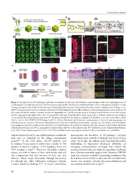
Figure 4. The application of bioprinting to fabricate vascularization and skin. (A) Schematic representation of the (i) bioprinting process of
a multilayered vascularized construct. (ii) Fluorescence micrographs showing the endothelialization of the void spaces (created as a result
of using permanent and sacrificial inks during the bioprinting process) and cross-sections showing a lumen. Adapted from Ouyang et al.,
2020 , licensed under Creative Commons Attribution 4.0 International License. (B) A strategy to promote angiogenesis based on the use of
[48]
(i) a sacrificial ink that renders a channel and (ii) cell-degradable ink. (iii) Microscopy analysis showing the endothelialization of the channel
and the angiogenic sprouting within the cell-degradable hydrogel. Magnifications show the presence of lumen structures (as small as
~10 µ) and the three-dimensional architecture . Reprinted (adapted) from Song KH, Highley CB, Rouff A, et al., Adv Funct Mater, 2018,
[49]
28(31):1–10. At 2018 WILEY-VCH Verlag GmbH Co. KGaA, Weinheim. (C) Schematic representation of the (i) bioprinting design of a
multilayered skin construct. (ii) Experimental program involving skin bioprinting, implantation, explantation, and analysis. (iii) Histological
micrographs showing the multilayered architecture and dermal markers of expression in human skin (for reference), grafted bioprinted skin
without placental pericytes (PCs), and grafted bioprinted skin with PCs in the dermal bioink. EC: Endothelial cells. Adapted from Baltazar
et al., 2020, Tissue Engineering Part A, 26: 5-6 . The publisher for this copyrighted material is Mary Ann Liebert, Inc. publishers.
[62]
critical situation faced by one million patients worldwide demonstrates the feasibility of 3D printing a full-size
each year, as estimated by the outage management human heart made entirely of alginate (not yet including
system . In the USA alone, 103,655 patients will cells). To do this, the authors used Freeform Reversible
[68]
be waiting for an organ by 2020; these include 91,790 Embedding of Suspended Hydrogels (FRESH), an
patients in need of a kidney, 12,521 needing a liver, and emerging extrusion-based technique that enables the
3504 needing a heart . These are all highly complex printing of practically any shape by injecting a hydrogel
[69]
organs, not only in terms of their architecture and cell into a thermo-reversible support bath. However, FRESH
type composition but also because of their size and (and any currently available bioprinting technique) has
function. Major organ fabrication through bioprinting its limitation in its ability to fabricate full-size functional
(or through any other fabrication technique) remains tissues. For example, printing this non-cellularized and
a major unsolved challenge [70,71] . A recent contribution non-structured heart took 4 days .
[71]
72 International Journal of Bioprinting (2021)–Volume 7, Issue 2