
Page 105 - IJB-7-4
P. 105
Ye Li, et al.
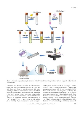
Figure 1. Schematic diagram depicting the fabrication of the 3D-printed β-tricalcium phosphate/poly lactic-co-glycolic acid carfilzomib-
loaded scaffolds.
P.R. China) was dissolved in 10 mL of dichloromethane solution) were dissolved in 200 μL of collagen I solution
(DCM) and vortex-stirred for 15 min until the PLGA was (9 mg/mL) at 4°C, and the CFZ/collagen I solution was
fully dissolved. Then, 1.8 mL of deionized (DI) water homogeneously mixed with TP inks to obtain CFZ/TCP/
and 50 µL of Tween 20 water were added to the PLGA PLGA/DCM emulsion ink (designated as “CTP”). The
to prepare 10 mL of PLGA organic solution. Afterward, as-formulated CTP inks were added into a 20 mL syringe
3 g of β-TCP powder mixture was mixed in the solution which was connected to a V-shaped plastic nozzle (inner
by ultrasonic vibration for 15 min to form TCP/DI water/ diameter: 0.4 mm) and further loaded into a cryogenic 3D
PLGA/DCM composite emulsion inks (designated as printer (Shenzhen Creality 3D Technology Co., Limited,
“TP”). To load CFZ in TP inks, 100 mg of CFZ and 4.6 P.R. China). Following the CAD file (a cylinder with a
μL of NaOH (1 N, to neutralize the acidic collagen I diameter of 4 mm and a height of 15 mm), porous 3D
International Journal of Bioprinting (2021)–Volume 7, Issue 4 101