
Page 109 - IJB-7-4
P. 109
Ye Li, et al.
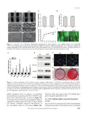
G H
A B
C D
I J
E F
Figure 2. Fabrication of a 3D-printed β-tricalcium phosphate/poly lactic-co-glycolic acid scaffold loaded with carfilzomib
(CFZ). (A-F) Scanning electron microscopy micrographs of different scaffolds at different magnification. (G) Compressive strengths of
cytidine triphosphate (CTP) scaffolds and TP controls. (H) Young modulus of the CTP scaffolds and TP controls. (I) Release behavior of
CFZ from CTP scaffolds in a 30-day test period. (J) Cell viability after seeding C3H10T1/2 in the scaffolds for 3 days.
A B C
D
Figure 3. Cytidine triphosphate (CTP) scaffolds promote osteogenic differentiation in C3H10T1/2 mesenchymal cells. (A) mRNA
expression of osteogenic genes in C3H10T1/2 cells stimulated with osteogenic medium supplemented with (carfilzomib [CFZ] group) or
without (control group) extracts of the CTP scaffold for 7 days. (B) Western blot results of osteogenic proteins in C3H10T1/2 cells from
control and CFZ group. (C) Immunofluorescence staining of osteocalcin in C3H10T1/2 cells from control and CFZ group. (D) Alizarin red
S staining of C3H10T1/2 cells stimulated with osteogenic medium supplemented with (CFZ group) or without (control group) extracts of
the CTP scaffold for 14 days. Scale bar = 50 μm. *P < 0.05.
mRNA expression levels. In addition, we performed Therefore, these data suggest that CTP scaffolds have
immunofluorescence staining of OCN in C3H10T1/2 cells good osteogenic capabilities in vitro.
from control and CFZ groups. As shown in Figure 3C,
OCN expression in CFZ group was remarkably enhanced 3.3. CTP scaffolds inhibit osteoclast formation
compared to control group. And within 14 days, alizarin in vitro
red staining confirmed enhanced mineralization of The role of CFZ in osteoclast activity has been mentioned
the extracellular matrix in CFZ group (Figure 3D). in the previous studies. In this study, we also investigated
International Journal of Bioprinting (2021)–Volume 7, Issue 4 105