
Page 112 - IJB-7-4
P. 112
CTP Scaffolds Treated Bone Defects
A E
B
C F
D
G
H
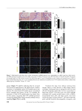
Figure 7. Histological results show that cytidine triphosphate scaffolds promote bone regeneration in a rabbit long bone defect model.
(A) H&E staining of the defect area at 12 weeks post-surgery. B: bone, F: Fibrous tissue, NB: New bone; V: Vessel). (B and E) IF analysis
and quantification of CTSK in bone sections from different groups. (C and F) Immunofluorescence analysis and quantification of CD31 in
bone sections from all groups. (D, G, and H) Immunofluorescence analysis and quantification of OCN and β-catenin in bone sections from
different groups. Scale bar = 50 μm. *P < 0.05.
groups (Figure 7C and F), indicating that the released Treatment for large bone defects resulting from
CFZ promoted angiogenesis in the defected area. Further, trauma, tumor, or other diseases is still a major clinical
expression of ontogenetic marker OCN and β-catenin was challenge. Autologous bone or autograft is still the most
detected. We noticed that OCN and β-catenin co-localized effective therapeutic approaches for bone regeneration in
in the cytoplasm and nucleus. In the CTP scaffolds, the large bone defect . In recent years, bone graft scaffolds
[23]
expression of both OCN and β-catenin was higher than combined with various growth factors, such as BMPs,
in other groups, suggesting that CTP scaffolds promoted mesenchymal stem cells, and other agents, have been
osteogenesis and possibly via the activation of the β-catenin used in the repair of bone defects . The most ideal bone
[24]
signaling (Figure 7D, G, and H). graft scaffold should be biocompatible, osteogenic, and
108 International Journal of Bioprinting (2021)–Volume 7, Issue 4