
Page 107 - IJB-7-4
P. 107
Ye Li, et al.
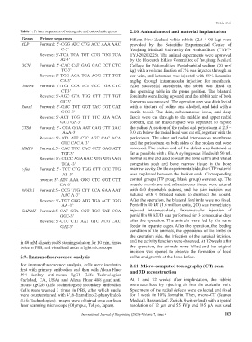
Table 1. Primer sequences of osteogenic and osteoclastic genes 2.10. Animal model and material implantation
Genes Primer sequences Fifteen New Zealand white rabbits (2.5 ± 0.5 kg) were
ALP Forward: 5’- CGG ATC CTG ACC AAA AAC provided by the Scientific Experimental Center of
C-3’ Youjiang Medical University for Nationalities (YYFY-
Reverse: 5’- TCA TGA TGT CCG TGG TCA TYJ-20200225). The animal experiments were approved
AT-3’ by the Research Ethics Committee of Youjiang Medical
OCN Forward: 5’- CAC CAT GAG GAC CCT CTC College for Nationalities. Pentobarbital sodium (20 mg/
TC-3’ kg) with a volume fraction of 3% was injected through an
Reverse: 5’- TGG ACA TGA AGG CTT TGT ear vein, and ketamine was injected with 50% ketamine
CA-3’ mg/kg through intramuscular injection for anesthesia.
Osterix Forward: 5’- TCT CCA TCT GCC TGA CTC After successful anesthesia, the rabbit was fixed on
CT-3’ the operating table in the prone position. The bilateral
Reverse: 5’- AGC GTA TGG CTT CTT TGT forelimbs were facing upward, and the rabbit hair of both
GC-3’ forearms was removed. The operation area was disinfected
Runx2 Forward: 5’- GAC TGT GGT TAC CGT CAT with a tincture of iodine and alcohol, and laid with a
GGC-3’ sterile towel. The skin, subcutaneous tissue, and deep
Reverse: 5’- ACT TGG TTT TTC ATA ACA fascia were cut through to the middle and upper radial
GCG GA-3’ forearm, and the muscle space was separated to expose
CTSK Forward: 5’- CCA GGA AAT GAG CTT GAC the radius. A section of the radius and periosteum at 2.5 –
AAA-3’ 3.0 cm below the radial head was cut off, together with the
Reverse: 5’- ATA ATT CTC AGT CAC ACA periosteum. The ulnar and radial interosseous membrane
GTC CAC A -3’ and the periosteum on both sides of the broken end were
MMP9 Forward:5’- CAC TCC CAC CCT GAG ATT removed. The broken end of the defect was flattened as
TGT-3’ far as possible with a file. A syringe was filled with 50 mL
Reverse: 5’- CCCC AGA GAC ATG ATG AAG normal saline and used to wash the bone debris and related
TCA -3’ congestion scab and bone marrow tissue in the bone
c-fos Forward: 5’- TGT CTG TGG CTT CCC TTG marrow cavity. On the experimental side, the CTP material
AT -3’ was implanted between the broken ends. Corresponding
reverse: 5’- ATC AAA GGG CTC GGT CTT control groups (TP group, blank group) were set up. The
CA -3’ muscle membrane and subcutaneous tissue were sutured
NFATc1 Forward:5’- CCG TTG CTT CCA GAA AAT with 4-0 absorbable sutures, and the skin incision was
AAC A -3’ sutured with 0 braided suture to disinfect the wound.
Reverse: 5’- TGT GGG ATG TGA ACT CGG After the operation, the bilateral forelimbs were not fixed.
AA -3’ Penicillin 40 kU (1.6 million units, QD) was immediately
GAPDH Forward:5’- CAT GTA CGT TGC TAT CCA injected intramuscularly. Intramuscular injection of
GGC-3’ penicillin 40 kU/D was performed for 3 consecutive days
Reverse: 5’- CTC CTT AAT GTC ACG CAC after the operation. The animals were fed by the same
GAT-3’ feeder in separate cages. After the operation, the feeding
condition of the animals, the appearance of the limbs on
the operation side, the infection of the surgical incision,
in 40 mM alizarin red S staining solution for 10 min, rinsed and the activity function were observed. At 12 weeks after
twice in PBS, and visualized under a light microscope. the operation, the animals were killed and the original
incision was opened to observe the formation of local
2.9. Immunofluorescence analysis callus and growth of the bone defect.
For immunofluorescence analysis, cells were incubated 2.11. Micro-computed tomography (CT) scan
first with primary antibodies and then with Alexa Fluor and 3D reconstruction
594 donkey anti-mouse IgG1 (Life Technologies,
Carlsbad, CA, USA) and Alexa Fluor 488 goat anti- At 8 and 12 weeks after implantation, the rabbits
mouse IgG2b (Life Technologies) secondary antibodies. were sacrificed by injecting air into the auricular vein.
Cells were washed 3 times in PBS, after which nuclei Specimens of the radial defects were collected and fixed
were counterstained with 4’,6-diamidino-2-phenylindole for 1 week in 10% formalin. Then, micro-CT (Scanco
(Life Technologies). Images were obtained on a confocal Medical, Bassersdorf, Zurich, Switzerland) with a spatial
laser scanning microscope (Olympus, Tokyo, Japan). resolution of 12 μm and 55 kVp and 145 μA was used
International Journal of Bioprinting (2021)–Volume 7, Issue 4 103