
Page 122 - IJB-7-4
P. 122
CNC-enhanced Hydrogels for 3D Bioprinting
A B C
D E F
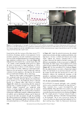
Figure 6. 3D printing results of hydrogels (20 wt% PCA +4.4 wt% cellulose nanocrystals). (A) Shear-thinning tests with the shear rate
2
from 0.01 to 100 rad/s. (B) Shear recovery experiments with alternating strain of 0.01% and 100% (black dot line). (C) The extrusion effect.
(D) Filament strength and stability with different size of nozzles. (E) The optical microscopic image of printed object and (F) The SEM
image of printed object after lyophilization.
linear decline with the increase of the shear rate at 37°C. in Figure 6E. Under the optical microscope, the printed
In addition, thixotropy, another critical parameter for the pattern showed relatively high resolution and fidelity with
printable estimation, was also conducted to measure the high layers (~4 mm), in which each filament was clearly
recovery of the mechanical properties of materials under a visible without any obvious extrusion-swell or even
large amplitude oscillatory force. The result (Figure 6B) collapse. Moreover, the details of printed constructs with
indicated that this material has a typical elastic effect (G’ different concentrations of inks are presented in Table S1.
> G’’) under a small amplitude strain (γ=0.01%). When Meanwhile, the SEM image of the lyophilized 3D printed
the strain is increased to a larger amplitude (γ=100%), pattern clearly exhibited the intact structure and framework
G’ decreases from 11 kPa to 3.6 kPa while G’’ increases (Figure 6F). For the unmodified PCA hydrogel (Figure
2
from 2.8 kPa to 4.0 kPa, exhibiting a quasi-liquid state. S1), the printed structure was totally collapsed and lumped
Moreover, the gel modulus varied reciprocally with the within 20 min based on our previous research . So far,
[16]
variation of strain amplitude, showing that this material the results illustrate that the introduction of CNCs can
had a favorable irreversibility, that is, thixotropy. effectively enhance the mechanical properties of the
Based on these results, extrusion-based 3D printing amorphous copolymers hydrogels, thereby significantly
was conducted using Bio-architect -PRO of Regenovo improving the extrudability and printability of the material.
®
for further evaluation. Through adjusting the temperature
in the cylinder to 37°C, the hydrogels could be extruded 3.5. In vitro cytotoxicity analysis
fluently as a filament rather than flowed as droplets
(Figure 6C), intuitively demonstrating that the enhanced PCLA-PEG-PCLA-formed hydrogels were originally
[40]
hydrogel has a required strength to be extruded. Then, widely used as injectable hydrogels for drug delivery .
filament collapse experiment was conducted using These materials show a good biocompatibility and
nozzles of syringe needles of different sizes for further biodegradability. Thus, fibroblasts were used to verify the
confirmation (Figure 6D). It is seen that different thickness former properties while lipase was used for the latter. The
of filaments could hanged straight on different gap distance enzymatic degradation properties were displayed in Figure
of scaffold, and no significant collapse can be observed, S2. The weight percent of hydrogel reduced linearly in
proving the favorable mechanical strength. By regulating 7-day incubation, showing a favorable biodegradability
the printing parameters (including size dimension, spacing of the hydrogels. The in vitro cytotoxicity during the
distance, and translational speed), the material could be 3-day cultivation was characterized and estimated by live/
extruded and printed as a 3D hydrogel construct, as shown dead staining assay. Under the fluorescent microscope
118 International Journal of Bioprinting (2021)–Volume 7, Issue 4