
Page 142 - IJB-7-4
P. 142
3D-Printed Anti-Tumor Scaffolds
A B
A
Figure 1. The morphology, composition, and structure of printed
polylactic acid/methotrexate (PLA/MTX) and PLA scaffolds. (A)
Scanning electron microscopy images of PLA/MTX scaffolds.
Scale bars represent 1 mm for A. Element mapping of C, N, and O
for the PLA/MTX scaffold. Scale bars represent 1 mm. (B) Energy-
dispersive spectrometer of porous printed PLA/MTX composite
scaffolds.
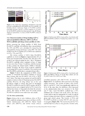
3.2. Characterization of drug loading (LE%) Figure 2. Methotrexate (MTX) release profiles of polylactic acid/
MTX (PLA/MTX) scaffolds of varying drug content over 7 and
and encapsulation efficiency (EE%) in PLA/ 30 days.
MTX scaffolds and drug release profiles in vitro
Before assessing the release profiles of MTX from
PLA/MTX scaffolds with different drug concentrations,
the LE% and EE% were calculated by measuring the
concentration of drug absorbance in the PBS. The EE%
of MTX was 98.1% and the LE% of MTX was controlled
at 0.5%, 1.5%, and 2.5%.
The release profiles of MTX from PLA/MTX
scaffolds are shown in Figure 2. Approximately 25% of
MTX was released within the first 24 h, and about 50%
of MTX was released within the first 7 days. 3D-printed
PLA/MTX scaffolds allow sustained release of drugs
in vitro more than 30 days. In addition, with the increase
of drug content, the release trend of MTX is basically
similar. Therefore, the drug release could be controlled
by adjusting MTX content in scaffolds. This result proved
that PLA/MTX scaffolds had a sustained release effect.
Figure 3 shows the comparison of MTX release Figure 3. Methotrexate (MTX) release profiles of polylactic acid/
profiles of 3D-printed PLA/MTX scaffolds of varying MTX (PLA/MTX) scaffolds of varying drug contents and cast
drug contents and cast PLA/MTX with 0.5% of MTX PLA/MTX with 0.5% of MTX over 5 days.
over 5 days. The drug release pattern of cast PLA/MTX
sample is similar to that of 3D-printed porous PLA/MTX osteoblast precursor cells (MC3T3-E1). As shown in
scaffold. However, the drug release rate of the cast Figure 4A-C, the inhibitory effect increases as the drug
PLA/MTX sample is slower than that of 3D-printed content increases. The growth of tumor cells (A549,
PLA/MTX scaffold. In cast PLA/MTX sample, the MTX MG-63, and MCF-7) has been significantly inhibited at
drug molecules were wrapped tightly by PLA material to 24 h. At the same time, the inhibitory effect increased
obstruct the release of drug molecules from the polymer as the administration time was extended to the third
matrix. The release of MTX from 3D-printed porous and 5 days. Each PLA/MTX scaffold has a 95%
th
PLA/MTX scaffold is more stable than cast sample. inhibition rate of tumor cells at the 120 h. The above
results indicate that the implantable PLA/MTX scaffold
3.3. In vitro cytotoxicity showed strong anti-tumor efficacy compared with PLA
Figure 4 shows the in vitro inhibitory effect of PLA/ group and control group. The toxicity of PLA and
MTX scaffolds on human lung cancer cell lines (A549), PLA/MTX scaffolds on normal MC3T3 cells was also
human osteosarcoma cells (MG-63), human breast investigated (Figure 4D). The results showed that PLA
cancer cells (MCF-7), and normal mouse embryo scaffold had little effect on the growth of normal cells,
138 International Journal of Bioprinting (2021)–Volume 7, Issue 4