
Page 144 - IJB-7-4
P. 144
3D-Printed Anti-Tumor Scaffolds
A
B C
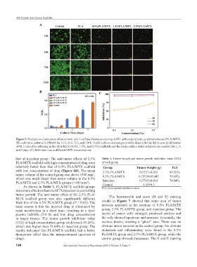
Figure 5. Evaluation of anticancer effect in vitro. (A) Live/Dead fluoresce staining of 4T1 cells on polylactic acid/methotrexate (PLA/MTX).
The cells were cultured in DMEM for 12 h, 24 h, 72 h, and 120 h. Viable cells are stained green while dead cells red. (B) In vitro proliferation
of 4T1 cells after culturing in the PLA/MTX (0.5%, 1.5%, and 2.5%) scaffolds and the tissue culture plates substrate (as control) for 1, 3,
and 5 days. (C) Inhibition rate at different MTX concentrations.
that of injection group. The anti-tumor effects of 2.5% Table 1. Tumor weight and tumor growth inhibition value (TGI)
PLA/MTX scaffold with high concentration of drug were of each group
relatively better than that of 0.5% PLA/MTX scaffold Group Tumor weight (g) TGI
with low concentration of drug (Figure 6D). The mean 2.5% PLA/MTX 0.1557±0.024 89.26%
tumor volume of the control group was above 1300 mm , 0.5% PLA/MTX 0.33524±0.042 76.88%
3
which was much larger than tumor volume in the 0.5% Injection 0.3763±0.018 74.04%
PLA/MTX and 2.5% PLA/MTX groups (<400 mm ). Control 1.45±0.3 -
3
As shown in Table 1, PLA/MTX scaffold groups TGI, tumor growth inhibition value.
were more effective than the MTX injection in controlling
tumor growth. The anti-tumor effect of the 2.5% PLA/ The hematoxylin and eosin (H and E) staining
MTX scaffold group was also significantly different
from that of the 0.5% PLA/MTX group (P < 0.05). The results in Figure 7 showed that some area of tumor
main reason is that the injected drug is eliminated by necrosis appeared in the sections of 0.5% PLA/MTX
renal metabolism in a short time, resulting in a short group, 2.5% PLA/MTX group, and injection group. The
plasma half-life (5-8 h) and low drug concentration nuclei of cancer cells enlarged, produced cavities and
in target tissues. The tumor growth inhibition value the cells showed apoptosis and necrosis. Eventually, the
(TGI) of high concentration group could reach 89.26%, nucleus breaks, creating a “ghost” area. There was no
which was higher than 74.04% in injection group. The obvious tumor necrosis in the control group. No obvious
results indicated that PLA/MTX scaffold had a better metastasis and inflammatory were found in the 0.5%
therapeutic effect than the intraperitoneal injection of PLA/MTX group and 2.5% PLA/MTX group, while the
drugs. control group showed metastases. The H and E staining
140 International Journal of Bioprinting (2021)–Volume 7, Issue 4