
Page 143 - IJB-7-4
P. 143
Mei, et al.
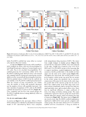
A B
C D
Figure 4. Evaluation of anticancer effect in vitro. In vitro proliferation of MCF-7(A), MG- 63 (B), A549 (C), and MC3T3 (D) cells after
culturing in the polylactic acid/methotrexate (0.5%, 1.5%, and 2.5%) scaffolds and the tissue culture plate substrate (as control) for 1, 3,
and 5 days.
while PLA/MTX scaffold had some effect on normal with intraperitoneal drug injection of MTX. The minor
cells, but the effect was limited. body weight changes in implant groups (Figure 6A)
As shown in Figure 5, the toxicity of PLA and PLA/ proved that the implanted scaffolds were well tolerated
MTX scaffolds on cancer cells was also investigated by by the mice. Weight loss occurred in the 0.5% PLA/
Live/Dead fluorescence staining of mouse breast cancer MTX and 2.5% PLA/MTX groups at the beginning due
4T1 cells. Due to its excellent biocompatibility, PLA to surgical anesthesia, surgical implantation, and other
scaffold group has low cytotoxicity within 120 h. The operations, but the growth status in the middle and later
PLA/MTX scaffold groups showed a lower cell survival stages was the same as the control group (Figure 6A).
rate compared with PLA group and control group, and the Throughout the experiment, the overall growth of mice
inhibitory effect increased with the prolonged culture time. in the negative control group was good, but one mouse
With the gradual release of MTX from the scaffold, the in the control group was paralyzed due to excessive
cell viability in the PLA/MTX scaffold group decreased. compression of the spine by the tumor. In the early stage,
With the increase of drug concentration and culture the growth state of the injection group was the same as
time, the number of living cells gradually decreased that of the control group. In the later stage, there were
and the number of dead cells gradually increased. The some adverse phenomena, such as weight loss, bad hair,
total induction of apoptosis of 4T1 cells by PLA/MTX small and sticky stool, and scorched yellow urine. There
scaffolds increased from 52.89% to 99.63% (Figure 4C). was no significant difference in organ coefficient of
Thus, compared with the PLA and control groups, the heart, liver, lung, and kidney among groups. The organ
PLA/MTX scaffolds showed strong inhibitory effect on coefficient of spleen in injection group was much lower
tumor 4T1 cells. than the other three groups, which was found at the time
of dissection (Figure 6B). The tumor sizes in Figure 6C
3.4. In vivo anti-tumor effect can directly show that PLA/MTX scaffold implantation
As shown in Figure 6, the anti-tumor effects of PLA/ significantly inhibited the tumor growth compared with
MTX scaffolds were evaluated by subcutaneous xenograft control group. The anti-tumor effects of 0.5% PLA/MTX
model of 4T1 tumor-bearing Balb/c mice compared scaffold with low concentration of drug are similar to
International Journal of Bioprinting (2021)–Volume 7, Issue 4 139