
Page 141 - IJB-7-4
P. 141
Mei, et al.
2.5. Drug loading capacity and drug release pain (10 mg/kg, gavage, q.d./2 days) and ceftriaxone
sodium (Dalian Meilun Biotechnology, China) to prevent
The 500 mg of PLA/MTX scaffolds were placed in a
beaker containing 3 mL CHCl to fully dissolve, and post-operative infection (20 mg/kg, tail vein injection,
q.d./2 days). The mice were housed under the standard
3
then, phosphate-buffered saline (PBS) was used to extract conditions.
the drug. The absorbance of the PBS was determined by
ultraviolet (UV) spectrophotometer at 305 nm. (2) In vivo antitumor efficacy and mechanism studies
The 500 mg of scaffold was placed in a 5 mL
centrifuge tube containing 3 mL PBS and then shaken at When tumor volume reached approximately 62.50 ±
3 (
37°C with a speed of 100 r/min. The absorbance of the 10 mm 5 mm×5 mm×5 mm), mice bearing 4T1 tumors
scaffolds was determined by UV spectrophotometer at were randomly assigned to four groups with five mice
305 nm and calculated by the following formula. in each group. The injection group was administered an
Loading efficiency % (LE%) = (Weight of intraperitoneal injection of drugs every 2 days of MTX at
encapsulated drug/total mass of the PLA/ MTX scaffold) 4 mg/kg. The PLA/MTX scaffolds printed from filament
×100% were implanted near the tumors. The tumor size and the
Entrapment efficiency % (EE%) = (Weight of weight of mice were recorded every day. The animals
encapsulated drug / Weight of total added drug) × 100%. were euthanized at the end point (21 days), and the
major organs (heart, liver, spleen, lung, and kidney) were
2.6. In vitro experiments collected for weighing to calculate organ coefficient and
(1) Cell lines then fixed in 4% paraformaldehyde for histopathological
examination. The animal research was approved by the
Mouse embryo osteoblast precursor cells (MC3T3-E1), Experimental Animal Ethics Committee of Yangzhou
human osteosarcoma cells (MG-63), human breast cancer University (NSFC2020-HXXY-4).
cells (MCF-7), human lung cancer cell lines (A549),
and mouse breast cancer cells (4T1) were purchased 2.8. Statistical analysis
from Shanghai Cell Bank of the Chinese Academy of All the experiments were performed in triplicate. Results
Sciences (Shanghai, China), and cultured in DMEM (or were expressed as mean ± standard deviation (SD).
RPMI) containing 10% FBS and 100 U/mL penicillin- Bonferroni post-test was performed to assess statistical
streptomycin.
significance. Statistical analysis was performed using
(2) Cytotoxicity assay non-parametric Kruskal–Wallis tests. P < 0.05 was
considered statistically significant.
Cells were cultured using 24-well plates with a density
of 1 × 10 cells/well. When the cells adhered and spread 3. Results
4
in plates, the scaffolds were immersed into the culture
medium. The CCK-8 assay was performed on days 1, 3.1. Morphological, composition, and structural
3, and 5. Briefly, the culture medium of the sample was analysis
replaced with 100 μL DMEM (RPMI) and incubated
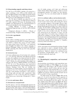
at 37°C for 2.5 h. The absorbance was measured at The morphology, microstructure, and compositions were
450 nm in the Multiskan FC enzyme labeling instrument investigated by SEM and EDS (Figure 1). According
(Thermo, Waltham, USA). The cell viability was to Figure 1A-1, SEM image of the PLA/MTX scaffold
evaluated by live/dead staining. After incubation in the showed a relatively smooth surface and had open big
medium for 12 h, 24 h, 72 h, and 120 h, 4 μM Calcein- pores in uniform size with integrated lines (755 ± 0.7 μm)
AM and 2 μM ethidium homodimer-1 were added to among the big pores. Moreover, the printed PLA/MTX
stain living cells and dead cells, respectively. After scaffolds were porous, with the porosity and pore size at
incubated at 37°C for 15 min, the cells were examined 29.7% and 309 ± 0.5 μm, respectively.
using fluorescence microscope (Olympusix53, Tokyo, The EDS spectra are shown in Figure 1B, and the
Japan). peaks of C, N, and O were observed in the spectrum of
PLA/MTX scaffold. The appearance of N peak confirmed
2.7. In vivo anti-tumor effect that MTX was introduced into PLA matrix. The color
map obtained from EDS analysis in Figure 1A shows
(1) Subcutaneous xenograft model the N (blue) distribution in the scaffold structure, which
The 4T1 cells (2 × 10 ) were subcutaneously injected into proves that MTX was well distributed in the PLA matrix.
5
the back of the BALB/c mice to establish the subcutaneous The evenly dispersion of MTX in the scaffold provides
xenograft model. After surgery, celecoxib (Dalian Meilun the possibility for MTX to be released along with PLA
Biotechnology, China) was given to relieve postoperative degradation.
International Journal of Bioprinting (2021)–Volume 7, Issue 4 137