
Page 55 - IJB-7-4
P. 55
Lin, et al.
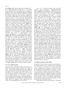
tubes (Figure 3B), and the compressive strength (9.67 – Ke et al. [101] prepared scaffolds with core-shell
26.72 MPa) and bending strength (15.21 – 21.12 MPa) structure using β-tricalcium phosphate/β-calcium
of the scaffold could be well controlled by changing the silicate (Figure 4A) with different combinations (CaSi @
parameters of the bone-mimicking structure to simulate CaP, CaP CaSi, CaSi, and CaP), and by adjusting the
@
the bone growth process, and the scaffold demonstrated composition distribution, it was found that CaSi CaP
@
the ability to induce new bone formation, angiogenesis showed a faster degradation rate within 7 – 14 days
and neurogenic differentiation and accelerate the growth (35%), while CaP CaSi microspheres showed excellent
@
of blood vessels in in vitro experiments, indicating that surface bioactivity and osteogenic activity (BV/TV,
multi-cellular delivery has great potential. Wang et al. 33%). Pistry et al. [102] used alginate gel or alginate/
[97]
prepared a porous β-tricalcium phosphate scaffolds with poly(ethylene glycol) diacrylate hybrid hydrogel as the
channel less, single-channel, and multi-channel structures scaffold shell in coaxial printing, and the core material
based on a single-layer cylindrical scaffold (Figure 3C), was separately used in three hydrogels encapsulating
which can achieve better cellular penetration and different cells (3T3-GelMA, HepG2-COL, and human
enhanced vascularization using interconnected channels umbilical vein endothelial cells [HUVEC]-Matrigel)
and pores in the scaffold to facilitate nutrient transport. (Figure 4B). The experimental results showed that
The macrostructure and microsurface topography of the the scaffold had good mechanical properties (elastic
implant play an important role in bone tissue regeneration. modulus up to 500 kPa) after the addition of PRGDA to
Chimene et al. developed a gelatin methacrylate the alginate gel, and the cells on the scaffold as a whole
[98]
(GelMA) slurry based on nanoengineered ionic- exhibited high biological activity, which remained above
covalent entanglement in a nut-like scaffold structure 90% after 28 days of in vitro culture. Taymour et al. [103]
(Figure 3D). With the increasing GelMA concentration, developed a core-shell structure scaffold using alginate
the compressive strength and toughness also increases and methylcellulose to loaded hepatocytes through a 3D
(103 kPa, 78 kJ/m 7.5 wt%). It also showed high extrusion-based bioprinting method (Figure 4C), which
3 (
printability, excellent enzymatic degradability (no more effectively constructed a microenvironment that allowed
than 20% degradation in 60 days), and osteoinductive coculture of hepatocytes with other liver-specific cells.
properties. Moon et al. designed 3D printed scaffolds Jin et al. prepared a calcium silicate core-shell structure
[99]
[92]
with hollow structures using alumina powder with a scaffold containing different mass fractions of Mg ions
particle size of 0.3 μm and camphene (Figure 3E), and by a coaxially aligned bilayer nozzle device (Figure 4D),
the scaffold had a compressive strength of approximately and the presence of Mg increased the compressive
5.4 MPa and a porosity of up to 86%, and the resulting strength of the scaffold from 39.4 MPa (CSi-Mg4) to 80
alumina filaments exhibited a highly microporous MPa (CSi-Mg10), and the degradation rate of CSi-Mg10
structure that could effectively stimulate cell-matter after 6 weeks was only 4.3%. Hong et al. [104] prepared a
interactions to induce new bone shapes. Ye et al. [100] gelatin-polyethylene glycol-tyrosamine-based core-shell
prepared hollow-structured gel scaffolds using GelMA structure based on a coaxial extrusion device using a one-
(Figure 3F) to assess biocompatibility and neuronal step gel bioprinting method (Figure 4E), and achieved
differentiation by culturing PC-12 cells and neural crest radial distribution of multiple vascular cells by loading
stem cells in vitro, and coculture experiments showed HUVECs with human dermal fibroblasts in tyramine,
that the average cell viability of nerve guidance conduits demonstrating that one-step generation of the idea of
with different inner diameters was 97.2%, 95.6%, and vascular structures is feasible.
95.1%, and close to 100%, respectively, and PC-12 cells
on GelMA scaffolds did not show any cytotoxic effects. 3.5. Bionic structures and others
3.4. Core-shell structure In addition to the four main structures mentioned above,
the overall structure of the scaffold can be customized
Adopting a physical structure similar to that of fiber optic to fit the shape of the defect or to simulate the organ
cables, the core-shell structure consists of two types of contour, depending on the skeletal defect and the need
slurry – internal and external, and is divided in different for functionality of the scaffold. For example, to repair
tubes and extruded in the form of a shell material closely bone defects more efficiently and precisely, designers try
covering the core material. Most of the common core- to print scaffolds into bionic structures such as meniscus-
shell structured supports are manufactured by coaxial shaped and ear-shaped. To address the defects in patients’
dual-jet printing devices. The core-shell structure is bodies more specifically, scaffolds are often designed into
characterized by the independence of the printing paste in special structures such as boat-shaped, spring-shaped,
the pre-printing process and inside the molded scaffold, and scroll-shaped, which can easily encapsulate cells and
which allows the scaffold to have degradability and growth factors or load other drugs [105] .
bioactivity with adjustable fast and slow rates. Inspired by the rosette structure, Han et al. [106] prepared
International Journal of Bioprinting (2021)–Volume 7, Issue 4 51